. 2021 May 12;13(5):892.
doi: 10.3390/v13050892.

PERK Is Critical for Alphavirus Nonstructural Protein Translation

Affiliations

PERK Is Critical for Alphavirus Nonstructural Protein Translation

Bibha Dahal et al. Viruses. .

Retraction in

Abstract

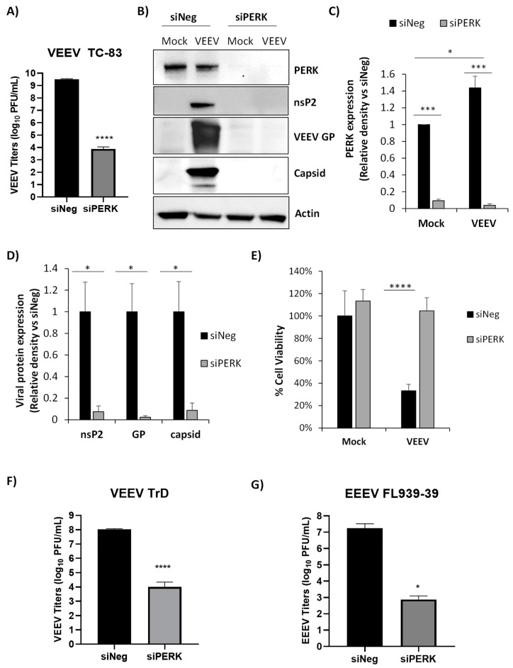
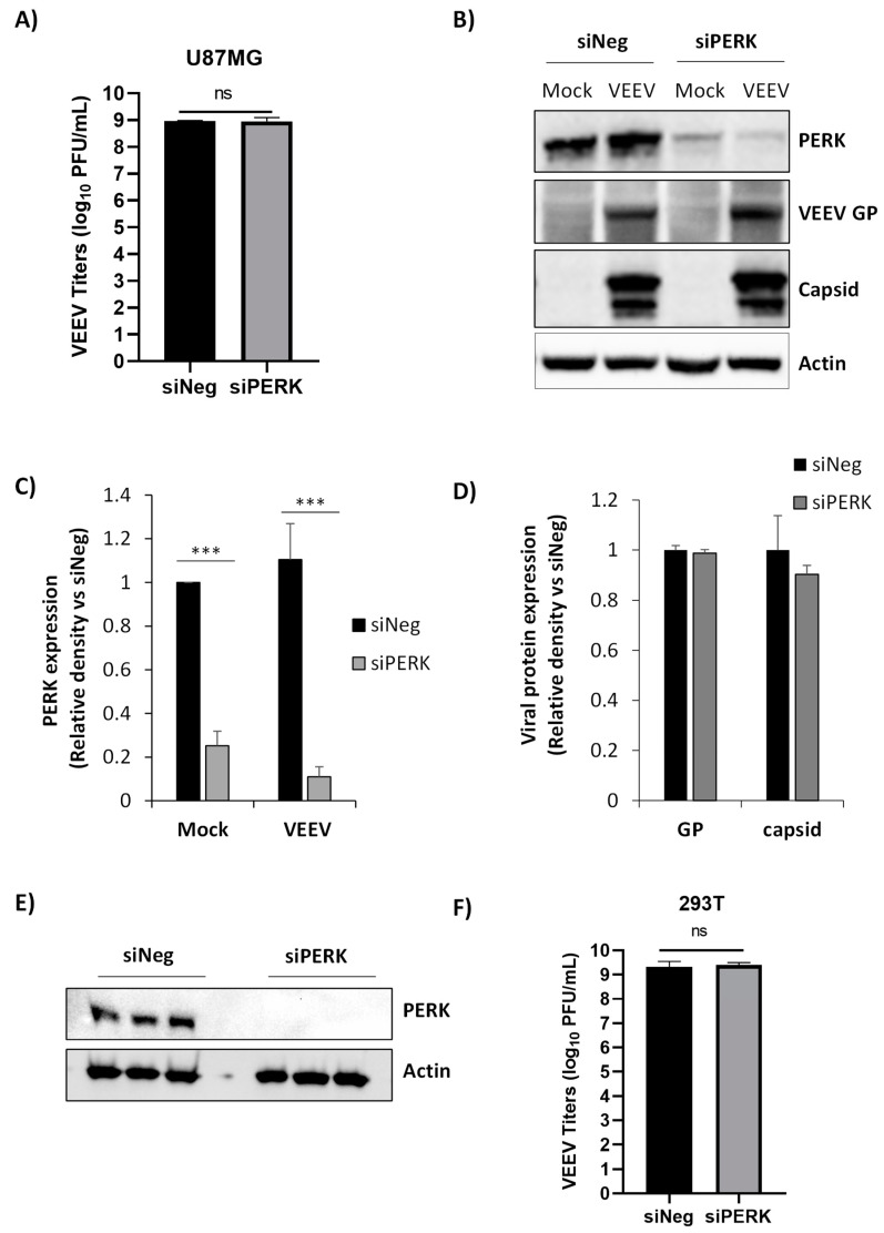
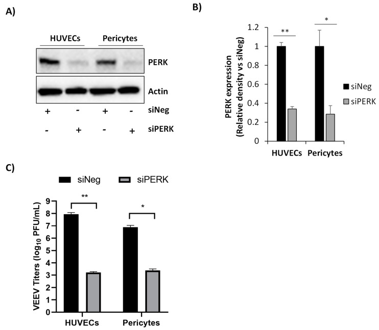
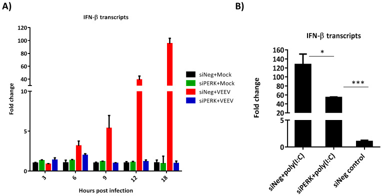
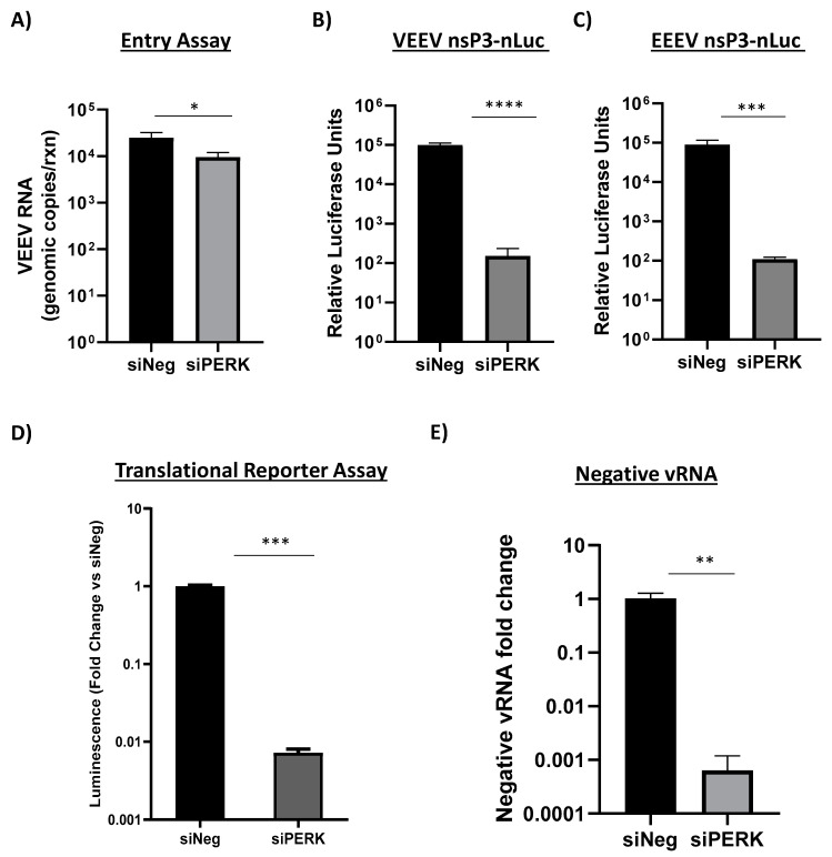
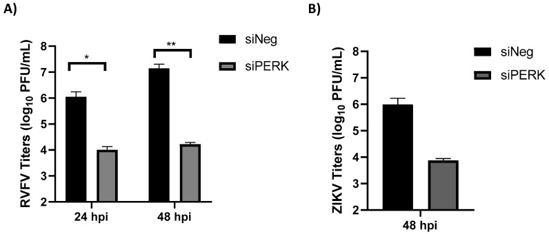
Venezuelan equine encephalitis virus (VEEV) is an alphavirus that causes encephalitis. Previous work indicated that VEEV infection induced early growth response 1 (EGR1) expression, leading to cell death via the protein kinase R (PKR)-like endoplasmic reticulum kinase (PERK) arm of the unfolded protein response (UPR) pathway. Loss of PERK prevented EGR1 induction and decreased VEEV-induced death. The results presented within show that loss of PERK in human primary astrocytes dramatically reduced VEEV and eastern equine encephalitis virus (EEEV) infectious titers by 4-5 log10. Loss of PERK also suppressed VEEV replication in primary human pericytes and human umbilical vein endothelial cells, but it had no impact on VEEV replication in transformed U87MG and 293T cells. A significant reduction in VEEV RNA levels was observed as early as 3 h post-infection, but viral entry assays indicated that the loss of PERK minimally impacted VEEV entry. In contrast, the loss of PERK resulted in a dramatic reduction in viral nonstructural protein translation and negative-strand viral RNA production. The loss of PERK also reduced the production of Rift Valley fever virus and Zika virus infectious titers. These data indicate that PERK is an essential factor for the translation of alphavirus nonstructural proteins and impacts multiple RNA viruses, making it an exciting target for antiviral development.

Keywords: PERK; Venezuelan equine encephalitis virus (VEEV); alphavirus; eastern equine encephalitis virus (EEEV); translation.


Conflict of interest statement

The authors declare no conflict of interest. The funders had no role in the design of the study; in the collection, analyses, or interpretation of data; in the writing of the manuscript, or in the decision to publish the results.

Figures

Figure 1
Figure 1
siRNA knockdown of protein kinase R (PKR)-like endoplasmic reticulum kinase (PERK) dramatically decreases Venezuelan equine encephalitis (VEEV) and eastern equine encephalitis virus (EEEV) replication in primary astrocytes. Primary human astrocytes were transfected with 100 nM of siNeg or siPERK siRNAs. At 48 h post transfection, cells were mock infected or infected with VEEV TC-83 (MOI 5). (A) Viral replication was analyzed via plaque assays in Vero cells using supernatants collected at 18 hpi. (B) Cell lysates were collected at 18 hpi and analyzed by immunoblot. PVDF membranes were probed for levels of PERK, VEEV nsP2, VEEV GP, and VEEV capsid. β-Actin was used as a loading control. (C,D) Data show the quantitation of the respective immunoblots. Protein levels expressed in each blot were normalized to β-actin and normalized values were calculated relative to siNeg-transfected cells. N = 3, * p ≤ 0.05, *** p ≤ 0.001. (E) Cell viability was measured using CellTiter-Glo assay at 48 hpi. Data were normalized to siNeg-transfected and mock-infected cells. Data are expressed as the mean  ±  SD (n  =  4). **** p ≤ 0.0001. (F,G) Primary astrocytes were transfected with 100 nM of siNeg or siPERK siRNAs. At 48 h post transfection, cells were infected with (F) VEEV TrD (MOI 5) or (G) EEEV FL93-939 (MOI 5). Viral replication was analyzed using supernatants collected at 18 hpi via plaque assays in Vero cells. Data are expressed as the mean  ±  SD (n  =  3 for siNeg and n = 5 for siPERK samples). * p ≤ 0.05, **** p ≤ 0.0001.
Figure 2
Figure 2
siRNA knockdown of PERK has no effect on VEEV replication in U87MG or 293T cells. U87MG cells were transfected with 100 nM of siNeg or siPERK siRNAs. At 48 h post transfection, cells were mock infected or infected with VEEV TC-83 (MOI 5). (A) Viral replication was analyzed via plaque assays using supernatants collected from U87MG cells at 16 hpi. N = 3, ns = not significant. (B) Cell lysates were collected at 16 hpi and analyzed by immunoblot. PVDF membranes were probed for levels of PERK, VEEV GP, and VEEV capsid. β-Actin was used as a loading control. (C,D) Data show the quantitation of the respective immunoblots. Protein levels expressed in each blot were normalized to β-actin and normalized values were calculated relative to siNeg-transfected cells. N = 3, *** p ≤ 0.001. (E) 293T cells were transfected with 100 nM of siNeg or siPERK siRNAs. At 48 h post transfection, cell lysates were collected and analyzed by immunoblot. PVDF membranes were probed for levels of PERK. β-Actin was used as a loading control. (F) 293T cells were transfected with 100 nM of siNeg or siPERK siRNAs. At 48 h post transfection, cells were infected with VEEV TC-83 (MOI 5). Viral replication was analyzed via plaque assays using supernatants collected at 18 hpi. N = 3, ns = not significant.
Figure 3
Figure 3
Loss of PERK decreases VEEV titers in human primary pericytes and human umbilical vein endothelial cells (HUVECs). (A) Pericytes or HUVECs were transfected with 100 nM of siNeg or siPERK siRNAs. Cell lysates were collected 48 h post transfection and analyzed by immunoblot. PVDF membranes were probed for levels of PERK. β-actin was used as a loading control. (B) Quantitative data of panel A. PERK protein levels were normalized to β-actin and normalized values were calculated relative to siNeg-transfected cells. (C) At 48 h post transfection, cells were infected with VEEV TC-83 (MOI 5), and viral replication was analyzed using supernatants collected at 18 hpi via plaque assays in Vero cells. Data are expressed as the mean  ±  SD (n  =  3). * p ≤ 0.05, ** p ≤ 0.01.
Figure 4
Figure 4
PERK impacts early events of VEEV replication. (A,B) Primary astrocytes were transfected with 100 nM of siNeg or siPERK siRNAs prior to VEEV TC-83 infection (MOI of 5). (A) Supernatants were collected at the indicated time points and viral titers were determined via plaque assay. (B) Cell lysates were collected at the indicated time points. RNA extraction was performed, and viral genomic copies were determined by RT-qPCR. Data are expressed as the mean  ±  SD (n  =  3). ** p ≤ 0.01, *** p ≤ 0.001, **** p ≤ 0.0001.
Figure 5
Figure 5
Loss of PERK does not result in elevation of interferon beta (IFN-β) production. Primary astrocytes were transfected with 100 nM of siNeg or siPERK siRNAs. At 48 h post transfection, (A) Cells were infected with VEEV TC-83 (MOI 5). Total RNA was isolated at 3, 6, 9, 12, and 18 hpi. IFN-β gene expression was determined by RT-qPCR. Fold changes were calculated relative to 18S ribosomal RNA and normalized to mock samples using the ΔΔCt method. Data are expressed as the mean  ±  SD (n  =  3). (B) Cells were treated with poly(I:C) complexed with Lipofectamine 2000 for 6 h. siNeg transfected cells treated with Lipofectamine only was used as a control. IFN-β gene expression determined as described above. Data are expressed as the mean  ±  SD (n  =  3). * p ≤ 0.5, *** p ≤ 0.001.
Figure 6
Figure 6
Loss of PERK signaling minimally impacts VEEV entry but inhibits the translation of incoming alphavirus genomes and production of negative-stranded viral RNA. (A) Primary astrocytes were transfected with 100 nM of siNeg or siPERK siRNAs. At 48 h post transfection, cells were infected with VEEV (MOI 5) and RNA collected 1 hpi. Viral genomic copies were determined by RT-qPCR. Results are displayed as genomic copies in logarithmic scale. Data are expressed as the mean  ±  SD (n  =  3). * p ≤ 0.05 using Student’s t-test. (B,C) Primary astrocytes were transfected with 100 nM of siNeg or siPERK siRNAs. At 48 h post transfection, cells were infected with VEEV nsP3-nLuc (panel B) or EEEV nsP3-nLuc at MOI of 5 (panel C). At 18 hpi, luminescence was measured using Promega’s Nano-Glo Luciferase Assay system. Data are expressed as the mean  ±  SD (n  =  5). *** p ≤ 0.001, **** p ≤ 0.0001. (D) Transfected cells were electroporated with translation reporter RNAs. Cells were lysed 2 h post electroporation and luciferase activity was measured. siPERK transfected and VEEV reporter RNA electroporated cells are expressed as RLUs (relative luminescence units) per µg protein expressed as a fold change over siNeg transfected and VEEV reporter RNA electroporated cells. Data are expressed as the mean  ±  SD (n  =  3). *** p ≤ 0.001. (E) Primary astrocytes were transfected with 100 nM of siNeg or siPERK siRNAs. At 48 h post transfection, cells were infected with VEEV TrD at MOI of 5. At 18 hpi, RNA was extracted and negative-stranded viral RNA detected via RT-qPCR. siNeg samples were set to a fold change of 1. Data are expressed as the mean  ±  SD (n  =  3). ** p ≤ 0.01.
Figure 7
Figure 7
Loss of PERK results in the reduction of Rift Valley fever virus (RVFV) and Zika virus (ZIKV) infectious titers. Primary human astrocytes were transfected with 100 nM of siNeg or siPERK siRNAs. At 48 h post transfection, cells were infected with (A) RVFV MP12 (MOI 5) or (B) ZIKV MR776 (MOI 5). Viral supernatants were collected at the indicated time points and viral titers determined via plaque assay. Data are expressed as the mean  ±  SD (n  =  3). * p ≤ 0.5, ** p ≤ 0.02.

Similar articles

Cited by

References

    1. Weaver S.C., Ferro C., Barrera R., Boshell J., Navarro J.-C. Venezuelan Equine Encephalitis. Annu. Rev. Entomol. 2004;49:141–174. doi: 10.1146/annurev.ento.49.061802.123422. - DOI - PubMed
    1. Sidwell R.W., Gebhardt L.P., Thorpe B.D. Epidemiological aspects of venezuelan equine encephalitis virus infections. Bacteriol. Rev. 1967;31:65–81. doi: 10.1128/BR.31.1.65-81.1967. - DOI - PMC - PubMed
    1. Gardner C.L., Burke C.W., Tesfay M.Z., Glass P.J., Klimstra W.B., Ryman K.D. Eastern and Venezuelan equine encephalitis viruses differ in their ability to infect dendritic cells and macrophages: Impact of altered cell tropism on pathogenesis. J. Virol. 2008;82:10634–10646. doi: 10.1128/JVI.01323-08. - DOI - PMC - PubMed
    1. Baer A., Lundberg L., Swales D., Waybright N., Pinkham C., Dinman J.D., Jacobs J.L., Kehn-Hall K. Venezuelan Equine Encephalitis Virus Induces Apoptosis through the Unfolded Protein Response Activation of EGR1. J. Virol. 2016;90:3558–3572. doi: 10.1128/JVI.02827-15. - DOI - PMC - PubMed
    1. Dahal B., Lin S.-C., Carey B.D., Jacobs J.L., Dinman J.D., van Hoek M.L., Adams A.A., Kehn-Hall K. EGR1 upregulation following Venezuelan equine encephalitis virus infection is regulated by ERK and PERK pathways contributing to cell death. Virology. 2019 doi: 10.1016/j.virol.2019.10.016. - DOI - PMC - PubMed

Publication types

MeSH terms

LinkOut - more resources