. 2021 May 12;13(5):894.
doi: 10.3390/v13050894.

Physical and Chemical Barriers in the Larval Midgut Confer Developmental Resistance to Virus Infection in Drosophila

Affiliations

Physical and Chemical Barriers in the Larval Midgut Confer Developmental Resistance to Virus Infection in Drosophila

Simon Villegas-Ospina et al. Viruses. .

Abstract

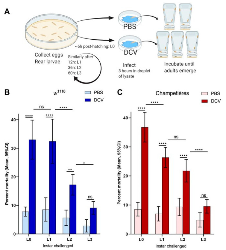
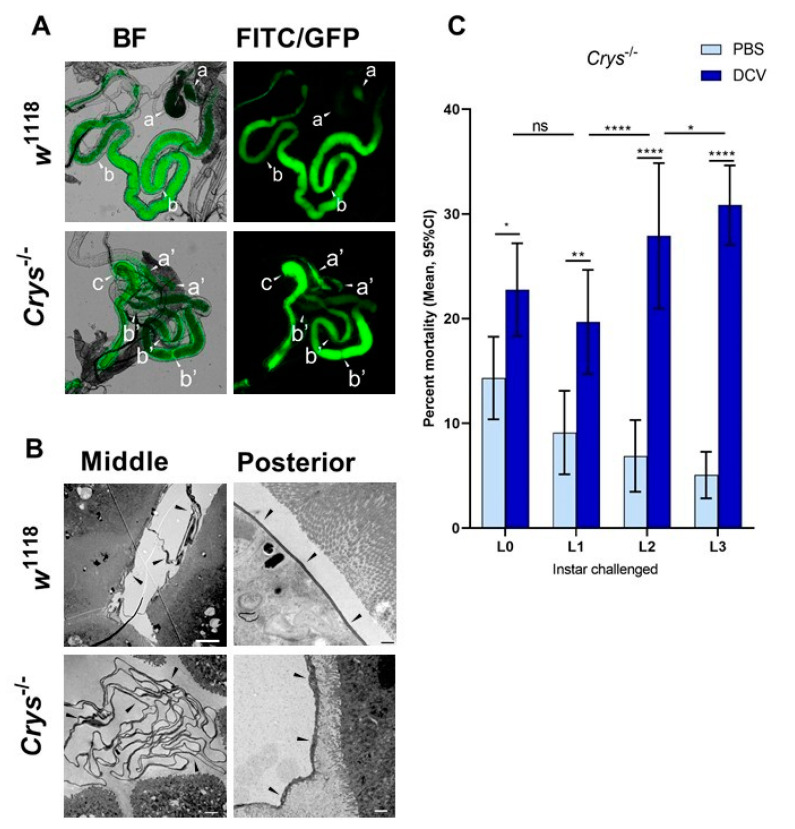
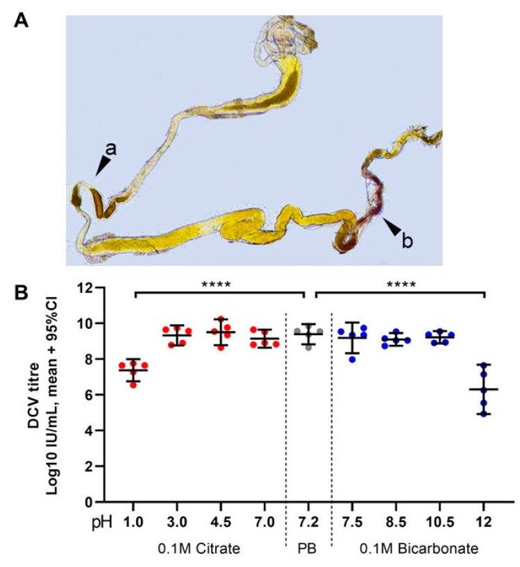
Insects can become lethally infected by the oral intake of a number of insect-specific viruses. Virus infection commonly occurs in larvae, given their active feeding behaviour; however, older larvae often become resistant to oral viral infections. To investigate mechanisms that contribute to resistance throughout the larval development, we orally challenged Drosophila larvae at different stages of their development with Drosophila C virus (DCV, Dicistroviridae). Here, we showed that DCV-induced mortality is highest when infection initiates early in larval development and decreases the later in development the infection occurs. We then evaluated the peritrophic matrix as an antiviral barrier within the gut using a Crystallin-deficient fly line (Crys-/-), whose PM is weakened and becomes more permeable to DCV-sized particles as the larva ages. This phenotype correlated with increasing mortality the later in development oral challenge occurred. Lastly, we tested in vitro the infectivity of DCV after incubation at pH conditions that may occur in the midgut. DCV virions were stable in a pH range between 3.0 and 10.5, but their infectivity decreased at least 100-fold below (1.0) and above (12.0) this range. We did not observe such acidic conditions in recently hatched larvae. We hypothesise that, in Drosophila larvae, the PM is essential for containing ingested virions separated from the gut epithelium, while highly acidic conditions inactivate the majority of the virions as they transit.

Keywords: Drosophila; antiviral mechanisms; dicistrovirus; gut pH; larval development; midgut; peritrophic matrix.


Conflict of interest statement

The authors declare no conflict of interest.

Figures

Figure 1
Figure 1
Mortality caused by DCV decreases when infection occurs later in larval development: larvae of the w1118 and Champetières fly lines were picked from rearing plates and orally challenged with lysates of DCV- or PBS-injected flies at 4 different times post ecclosion, and transferred to individual test plates (A). At least 4 biological replicates were done, each containing 2-PBS- and 3-DCV-challenged groups of larvae, per time point. Mortality was calculated from the proportion of adults that emerged from the larvae transferred to each test plate, for each PBS or DCV feeding challenge. A two-way ANOVA analysis was used to compare the effect of infection and the time (of larval development) at which the challenge was done. Using Šídák’s test for multiple comparisons, the survival between PBS and DCV at each time point was compared, to determine that L0, L2, and L2 larvae were susceptible to lethal DCV infection compared to their uninfected controls; and Tukey’s test was used to compare the mortality after PBS or DCV feeding across instars, to determine that the mortality caused by DCV decreased significantly between the L0-L1, L1-L2, and/or L2-L3 times of infection ((B,C); statistical analysis results are detailed in Table S1; * p < 0.05, ** p < 0.01, *** p < 0.001, **** p < 0.0001).
Figure 2
Figure 2
The size of the gut increases in a proportional manner to the total body size: larvae of the w1118 (blue circles: (A,C)) and Champetières (red circles: (B,D)), fly lines of different sizes and instars (L1, L2, and L3) were simultaneously selected and fed with standard solutions of FITC-dextran particles. Groups of 5 similarly sized larvae were weighed, surface cleaned, and macerated, to measure the ingested FITC content and estimate the volume of the gut. The correlation between average weight and average gut volume per group of 5 larvae was estimated using a Pearson correlation test (A,B), indicating a positive strong correlation between total body and gut size as the larvae grow. Furthermore, the gut-to-body ratio was calculated, and a Pearson test was used to evaluate its correlation to body size (C,D), indicating no correlation for Champetières, and a weak negative correlation for w1118 larvae.
Figure 3
Figure 3
The loss of Crys increases the susceptibility to oral viral infection in adult flies: (A) the Crys−/− line originates from a 7.8 kb Minos-mediated insertion within intron 1 of the Crys gene, Mi{ET1} [61,62]. Primers for Crys used in this study were designed for differentially amplifying the Mi{ET1} insertion, the wild-type Crys gene, and cDNA obtained from processed mRNA. (B) The insertion was confirmed by conventional PCR (1: 100bp DNA ladder; 2, 3, 4: wild-type Crys gene in w1118, Champetieres, and Crys−/−, respectively; 5, 6, 7: CrysMB08319 in w1118, Champetières, and Crys−/−, respectively). (C) A decreased or total loss of mRNA expression [24] was confirmed by RT-qPCR in pools of wandering larvae, pupae, or 1-day-old adults of w1118 and Crys−/−, and ‘nd’ signifies none-detected. Relative expression of Crys to RpL32 was analysed by ΔΔCt of the estimated efficiency for each primer, each previously calculated from the dilution series of a known, positive sample. (D) Adult males and females of 3–7 days-old were briefly starved and orally challenged with PBS- or DCV-lysates mixed with sucrose, and used for RNA extractions and survival bioassays. (E) Neither the starvation nor 6 h after having initiated the oral challenge (hours post-feeding, hpf) led to significant differences in the expression of Crys mRNA of the wild-type controls [24] when analysed by a two-way ANOVA using Šídák’s multiple comparisons; however, the expression was significantly higher in males than in females at every condition. No Crys mRNA was detected in Crys−/− adults at any time point of the oral feeding setup (data not shown). (F,G) After the feeding challenge, we tracked the survival of adults for 30 days. Analysis of survival data indicates that the loss of Crys significantly increases the mortality of DCV-fed adults, after the analysis of survival curves using a Cox mixed-effects model (*** p < 0.001; **** p < 0.0001).
Figure 4
Figure 4
The loss of Crys increases the permeability of the larval PM, and increases the mortality of older larvae after oral DCV infection: (A) using bright field (BF) and fluorescence microscopy, the crossing of FITC-labelled DCV-sized particles out of the PM and into the gut lumen was observed more commonly in the middle midgut of Crys−/− larvae than in their w1118 counterparts (pointers a, a’). In contrast, the PM of the posterior midgut was permeable to DCV-sized particles in almost every gut analysed (pointers b, b’). The transgenic 3xP3-EGFP marker in Crys−/− larvae was observed in the same fluorescence channel used for FITC, and used to confirm the Mi{ET1} cassette insertion. The promoter of this marker is reported to be brain- and eye-specific [64]; however, it also produces a signal in the hindgut (pointer c). (B) Using TEM, we evaluated the thickness and structure of the PM (indicated by the pointers) in middle and posterior midguts of L3 larvae of w1118 and Crys−/− larvae. In Crys−/−, the PM appeared diffuse, irregular, and tended to fold and accumulate in excess in the middle midgut. Bar in w1118-middle, 5 µm; in w1118-posterior, 0.5 µm; in both Crys−/− images, 1 µm. (C) Upon oral challenge following the same protocol as in Figure 1A, we used a two-way ANOVA with Šídák’s test to compare PBS and DCV mortality at each time point. At all time points there was an increased mortality when larvae were fed with DCV lysates. Furthermore, after comparing the effect of PBS or DCV across time points using Tukey’s test, the mortality caused by DCV significantly increased when the oral challenge was done in late larvae (results of statistical analysis are detailed in Table S1; * p < 0.05, ** p < 0.01, **** p < 0.0001).
Figure 5
Figure 5
Highly acidic and highly alkaline conditions found in the midgut can affect DCV infectivity: (A) upon dissection of L1, L2, and L3 larvae, the m-cresol purple ingested shifts to a red/orange hue in the middle midgut indicating a pH between 1.2 and 2.5 (pointer a), and to a brown/purple hue towards the end of the posterior midgut, close to the Malpighian tubules, indicating a pH > 9.0 (pointer b). These colours fade towards yellow rapidly after dissection, as the gut is neutralised when exposed to the glycerol-PBS. Hence, colours depicted do not accurately reflect those observed in vivo or during dissection; saturation was increased for illustrative purposes. In contrast, the guts of L0 did not display the red or purple colouration of m-cresol purple at acid (pH < 2.8) or alkaline (pH > 8.5) conditions, respectively, either before or after dissection (data not shown [48]). (B) Aliquots of purified DCV (stock at 2.5 × 109 = 109.4 IU/mL) were incubated under acidic-to-neutral (citrate; left red circles), neutral (PB control; centre grey circles), or neutral-to-alkaline (bicarbonate; right blue circles) buffers for 15 min at room temperature and used to infect Drosophila S2 cells. DCV titre was calculated by TCID50, and normalised using log-transformation. Each condition was compared to the PB-treated control (in which the purified DCV aliquots were maintained) using Šídák’s multiple comparisons test. Compared to the PB pH 7.2 treatment, highly acidic conditions (Citrate, pH 1.0) decreased the viral titre by 2 orders of magnitude (mean, 107.37 vs. 109.39 IU/mL), whereas highly alkaline conditions (bicarbonate, pH = 12.0) decreased the viral titre by 3 orders of magnitude (mean, 106.30 vs. 109.39 IU/mL) (p < 0.0001 for both comparisons). Acidic conditions found within the middle midgut (pH 1–2) may significantly decrease the DCV infectivity, whereas alkaline conditions similar to those found in the posterior midgut (pH 7.5–10.5) are likely not to affect the stability or the remaining infectious viruses.

Similar articles

Cited by

References

    1. Marques J.T., Imler J.-L. The diversity of insect antiviral immunity: Insights from viruses. Curr. Opin. Microbiol. 2016;32:71–76. doi: 10.1016/j.mib.2016.05.002. - DOI - PMC - PubMed
    1. Palmer W., Varghese F., van Rij R. Natural variation in resistance to virus infection in Dipteran insects. Viruses. 2018;10:118. doi: 10.3390/v10030118. - DOI - PMC - PubMed
    1. Evans H.F. The influence of larval maturation on responses of Mamestra brassicae L. (Lepidoptera: Noctuidae) to nuclear polyhedrosis virus infection. Arch. Virol. 1983;75:163–170. doi: 10.1007/BF01315270. - DOI - PubMed
    1. Li S.Y., Skinner A.C. Influence of larval stage and virus inoculum on virus yield in insect host Neodiprion abietis (Hymenoptera: Diprionidae) J. Econ. Entomol. 2005;98:1876–1879. doi: 10.1093/jee/98.6.1876. - DOI - PubMed
    1. Engelhard E.K., Volkman L.E. Developmental resistance in fourth instar Trichoplusia ni orally inoculated with Autographa californica M nuclear polyhedrosis virus. Virology. 1995;209:384–389. doi: 10.1006/viro.1995.1270. - DOI - PubMed

Publication types

LinkOut - more resources