. 2021 May 12;22(10):5114.
doi: 10.3390/ijms22105114.

Transient Global Ischemia-Induced Brain Inflammatory Cascades Attenuated by Targeted Temperature Management

Affiliations

Transient Global Ischemia-Induced Brain Inflammatory Cascades Attenuated by Targeted Temperature Management

Dae Ki Hong et al. Int J Mol Sci. .

Abstract

Sudden cardiac arrest leads to a significantly increased risk of severe neurological impairment and higher mortality rates in survivors due to global brain tissue injury caused by prolonged whole-body ischemia and reperfusion. The brain undergoes various deleterious cascading events. Among these damaging mechanisms, neuroinflammation plays an especially crucial role in the exacerbation of brain damage. Clinical guidelines indicate that 33 °C and 36 °C are both beneficial for targeted temperature management (TTM) after cardiac arrest. To clarify the mechanistic relationship between TTM and inflammation in transient global ischemia (TGI) and determine whether 36 °C produces a neuroprotective effect comparable to 33 °C, we performed an experiment using a rat model. We found that TTM at 36 °C and at 33 °C attenuated neuronal cell death and apoptosis, with significant improvements in behavioral function that lasted for up to 72 h. TTM at 33 °C and 36 °C suppressed the propagation of inflammation including the release of high mobility group box 1 from damaged cells, the activation and polarization of the microglia, and the excessive release of activated microglia-induced inflammatory cytokines. There were equal neuroprotective effects for TTM at 36 °C and 33 °C. In addition, hypothermic complications and should be considered safe and effective after cardiac arrest.

Keywords: apoptosis; high mobility box protein 1; inflammation; microglia; post cardiac arrest care; targeted temperature management.


Conflict of interest statement

The authors declare no conflict of interest.

Figures

Figure 1
Figure 1
Target temperature management (TTM) suppresses transient global ischemia (TGI)-induced neuronal death and sensorimotor deficit. (A) Brain sections were stained with Fluoro-Jade B fluorescent dye to detect degenerating neurons. TGI at a normal temperature of 37.5 °C significantly increased the number of fluorescent cells in the hippocampal cornus ammonis 1 region. These cells were significantly decreased after TGI-TTM at 33 °C and 36 °C. There were no differences between the sham groups. Scale bar = 50 μm. (B) Bar graph represents the total number of Fluoro-Jade B-positive cells (one way-ANOVA, multiple comparison following Bonferroni adjustment. Sham + 33 °C, n = 5; Sham + 36 °C, n = 5; Sham + 37.5 °C, n = 5; TGI + 33 °C, n = 8; TGI + 36 °C, n = 5; TGI + 37.5, n = 8. * p and # p < 0.05). (C) Adhesive removal test to observe sensorimotor deficit proceeded 3 to 72 h after TGI (tested daily). There were no differences in the removal time in each sham group assessment. TGI-TTM at 33 °C and 36 °C significantly reduced the removal time (time-dependent) compared to TGI at normal temperature at 37.5 °C (one way-ANOVA, multiple comparison following Bonferroni adjustment. Sham + 33 °C, n = 5; Sham + 36 °C, n = 5; Sham + 37.5 °C, n = 5; TGI + 33 °C, n = 9; TGI + 36 °C, n = 7; TGI + 37.5, n = 8. * p and # p < 0.05).
Figure 2
Figure 2
TTM suppresses apoptotic cell death 72 h after TGI. (A) Representative histological images display the number of TUNEL-positive cells in the cornus ammonis 1 of the sham or the hippocampus and in TGI. There were no differences in the number of TUNEL-positive cells in each sham group. TGI at a normal temperature at 37.5 °C resulted in an increase in the number of these cells at 72 h. TGI-TTM at 33 °C and 36 °C significantly decreased TUNEL-positive cells compared to TGI at normal temperature at 37.5 °C. Scale bar = 50 μm. (B) Representative bar graphs show the number of TUNEL-positive cells in the sham and TGI groups (one way-ANOVA, multiple comparison following Bonferroni adjustment. Sham + 33 °C, n = 5; Sham + 36 °C, n = 5; Sham + 37.5 °C, n = 5; TGI + 33 °C, n = 8; TGI + 36 °C, n = 5; TGI + 37.5, n = 8. * p and # p < 0.05). TTM at 33 °C and 36 °C reduces apoptosis-associated caspase-8 and Bcl-2-associated protein (BAX) expression at 72 h after TGI. The expression of apoptotic cell death-related caspase-8 and pro-apoptotic member, BAX, was suppressed by TTM at 33 °C and 36 °C after TGI. (C) In the sham groups, there were no differences in caspase-8 expression. TGI-normal temperature (NT) at 37.5 °C upregulated the expression of caspase-8 within the hippocampal cornus ammonis 1 region. TTM at 33 °C and 36 °C significantly suppressed caspase-8 expression. Scale bar = 50 μm. (E) The apoptosis-related substrate, BAX, was overexpressed in TGI-NT 37.5 °C. In the sham groups, there were no differences in BAX expression. TGI-TTM at 33 °C and 36 °C significantly reduced BAX expression. Scale bar = 50 μm. (D) Bar graph indicates the percentage of caspase-8 expression. Caspase-8 expression is greater in TGI-NT at 37.5 °C than at 33 °C and 36 °C (one way-ANOVA, multiple comparison following Bonferroni adjustment. Sham + 33 °C, n = 3; Sham + 36 °C, n = 3; Sham + 37.5 °C, n = 3; TGI + 33 °C, n = 3; TGI + 36 °C, n = 3; TGI + 37.5, n = 4. * p and # p < 0.05). (F) Graph shows that BAX expression decreased in both hypothermic paradigms (one way-ANOVA, multiple comparison following Bonferroni adjustment. Sham + 33 °C, n = 3; Sham + 36 °C, n = 3; Sham + 37.5 °C, n = 3; TGI + 33 °C, n = 3; TGI + 36 °C, n = 3; TGI + 37.5, n = 4. * p and # p < 0.05).
Figure 3
Figure 3
TTM blocks high mobility group box 1 (HMGB1) and cytokines release after TGI. HMGB1 level measured at 4 h after TGI. (A,B) Representative images showed co-localization of HMGB1 with nuclei (DAPI fluorescent stain). In the sham groups, HMGB1 expression was correlated with nuclei. TGI at normal temperature (37.5 °C) accelerated HMGB1 release from the nuclei, and TGI-TTM at 33 °C and 36 °C prevented HMGB1 release in the hippocampal cornus ammonis 1 region. Scale bar = 50 μm. (C,D) Percentage of HMGB1 and nuclei (DAPI) correlation in both hippocampal cornus ammonis 1 and parietal cortex in both hypothermic paradigms (one way-ANOVA, multiple comparison following Bonferroni adjustment. Sham + 33 °C, n = 4; Sham + 36 °C, n = 4; Sham + 37.5 °C, n = 4; TGI + 33 °C, n = 4; TGI + 36 °C, n = 4; TGI + 37.5, n = 4. * p and # p < 0.05). HMGB1 release from nuclei triggered pro-inflammatory cytokine expression. (EG) Quantification of fold change (2−ddct value) by quantitative polymerase chain reaction of tumor necrosis factor-alpha, interleukin-1β, and interleukin-6 versus glyceraldehyde 3-phosphate dehydrogenase. The expression of tumor necrosis factor-alpha, interleukin-1β, and interleukin-6 was decreased after TTM at 33 °C and 36 °C compared to the expression at 37.5 °C (normal temperature). TGI-TTM at 36 °C decreased tumor necrosis factor-alpha expression significantly more than TGI at 37.5 °C (one way-ANOVA, multiple comparison following Bonferroni adjustment. Sham, n = 3; TGI + 33 °C, n = 3; TGI + 36 °C, n = 3; TGI + 37.5, n = 3. * p and # p < 0.05).
Figure 4
Figure 4
TGI-induced high mobility group box 1 release in the early stage triggers microglial activation and M1 type polarization. TTM at 33 °C and 36 °C inhibits this. (A,B) At 72 h after TGI, representative images show microglial activation and M1 type polarization in both the sham and TGI groups in the hippocampal cornus ammonis 1 and parietal cortex. The sham groups had no differences in microglial activation and polarization. TGI to normal temperature at 37.5 °C triggered microglial activation (Iba-1, green signal) and M1 type polarization (CD86, red signal). Scale bar = 50 μm. (C,D) Image shows low and high magnification of Iba-1-positive microglial cells in hippocampal cornus ammonis 1 region and parietal cortex. Scale bar = 20 μm. (E,F) Bar graph shows the percentage of Iba-1 and CD86 emerging in the hippocampal cornus ammonis 1 and parietal cortex. TGI-TTM at 33 °C and 36 °C had a lower percentage than TGI at 37.5 °C (one way-ANOVA, multiple comparison following Bonferroni adjustment. Sham + 33 °C, n = 3; Sham + 36 °C, n = 4; Sham + 37.5 °C, n = 6; TGI + 33 °C, n = 5; TGI + 36 °C, n = 8; TGI + 37.5, n = 6. * p and # p < 0.05).
Figure 5
Figure 5
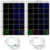
TTM at 33 °C and 36 °C suppresses changes in M2 phenotype microglia at 72 h after TGI. (A,B) Fluorescent imaging shows total microglia (Iba-1, green signal), the M2 type microglia (CD206, red signal), and merger with nuclei in the hippocampal cornus ammonis and parietal cortex regions. Scale bar = 50 μm. (C,D) Percentage of Iba-1 and CD206 merging at the hippocampal cornus ammonis and parietal cortex regions. Iba-1 and CD206 co-localization percentage was lower in TGI-TTM at 33 °C and 36 °C than in TGI at 37.5 °C (one way-ANOVA, multiple comparison following Bonferroni adjustment. Sham + 33 °C, n = 3; Sham + 36 °C, n = 4; Sham + 37.5 °C, n = 6; TGI + 33 °C, n = 5; TGI + 36 °C, n = 8; TGI + 37.5, n = 6. * p and # p < 0.05).
Figure 6
Figure 6
Cytokine array screening of the normal temperature and hypothermic paradigms of TGI. (AD) Representative chemiluminescent images show the proteome profiler array at normal temperature and TTM conditions at 24 h after TGI. Selected cytokines involving ciliary neurotrophic factor, fractalkine/CX3CL1 (chemokine ligand 1), slCAM-1/CD54, thymus chemokine/CXCL7 (chemokine ligand 7), and tissue inhibitor of metalloproteinases 1 are indicated in the box, and compared with sham or TGI to normal temperature at 37.5 °C, TTM at 33 °C and 36 °C. (EG) Analyzed value of the selected cytokines (fractalkine/CX3CL1, slCAM-1/CD54, tissue inhibitor of metalloproteinases 1) displayed by a representative bar graph. Expression of these cytokines was remarkably attenuated by TTM at 33 °C and 36 °C after TGI (one way-ANOVA, multiple comparison following Bonferroni adjustment. Sham, n = 3; TGI + 33 °C, n = 3; TGI + 36 °C, n = 3; TGI + 37.5, n = 3. * p and # p < 0.05).
Figure 7
Figure 7
Experimental design for targeted temperature management (TTM). (A) Timeline representing the entire experimental design following TTM. After termination of TTM, several analyses were conducted to verify the effects of TTM. (B) Changes of systemic body temperature during TTM. (CE) Arterial blood pressure and electroencephalograph pattern during transient global ischemia. Before initiation of the set isoelectric point; blood withdraw range 40 (diastolic) to 50 (systolic) mmHg. Blood pressure and electroencephalography findings were completely restored after blood reperfusion. Targeted temperature management (TTM) at 33 °C and 36 °C decreases hippocampal neuronal death and sensorimotor deficits.

References

    1. Donnino M.W., Andersen L.W., Berg K.M., Reynolds J.C., Nolan J.P., Morley P.T., Lang E., Cocchi M.N., Xanthos T., Callaway C.W., et al. Temperature management after cardiac arrest: An advisory statement by the advanced life support task force of the international liaison committee on resuscitation and the american heart association emergency cardiovascular care committee and the council on cardiopulmonary, critical care, perioperative and resuscitation. Circulation. 2015;132:2448–2456. doi: 10.1161/cir.0000000000000313. - DOI - PubMed
    1. Girotra S., Chan P.S., Bradley S.M. Post-resuscitation care following out-of-hospital and in-hospital cardiac arrest. Heart. 2015;101:1943–1949. doi: 10.1136/heartjnl-2015-307450. - DOI - PMC - PubMed
    1. Kang Y. Management of post-cardiac arrest syndrome. Acute Crit. Care. 2019;34:173–178. doi: 10.4266/acc.2019.00654. - DOI - PMC - PubMed
    1. Neumar R.W. Molecular mechanisms of ischemic neuronal injury. Ann. Emerg. Med. 2000;36:483–506. doi: 10.1016/S0196-0644(00)82028-4. - DOI - PubMed
    1. Yenari M.A., Han H.S. Neuroprotective mechanisms of hypothermia in brain ischaemia. Nat. Rev. Neurosci. 2012;13:267–278. doi: 10.1038/nrn3174. - DOI - PubMed