. 2021 May 12;13(10):2322.
doi: 10.3390/cancers13102322.

Utility and Mechanism of SHetA2 and Paclitaxel for Treatment of Endometrial Cancer

Affiliations

Utility and Mechanism of SHetA2 and Paclitaxel for Treatment of Endometrial Cancer

Vishal Chandra et al. Cancers (Basel). .

Abstract

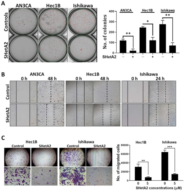
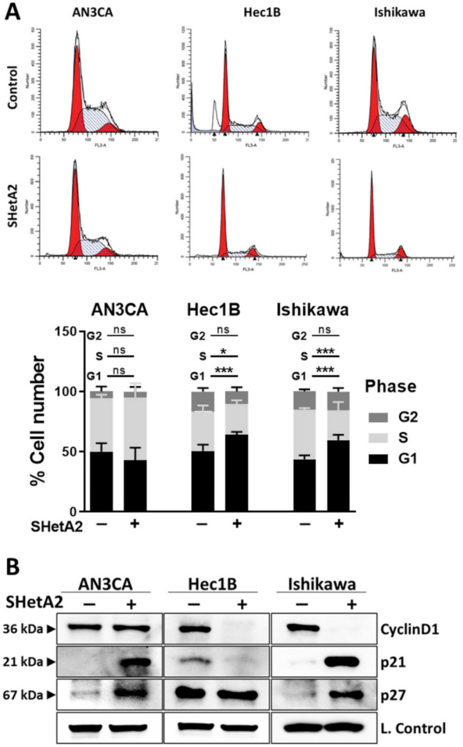
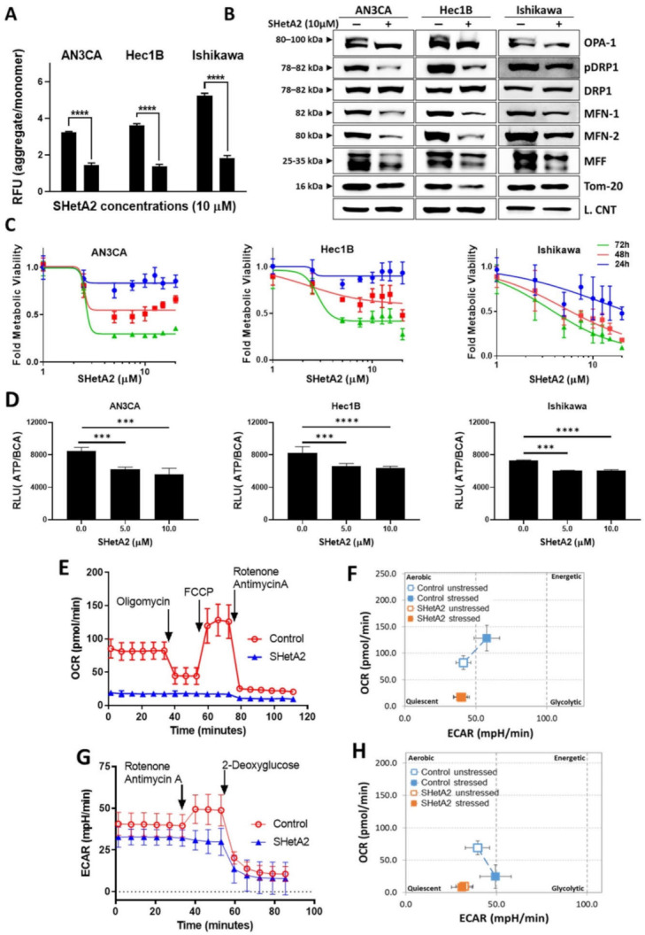
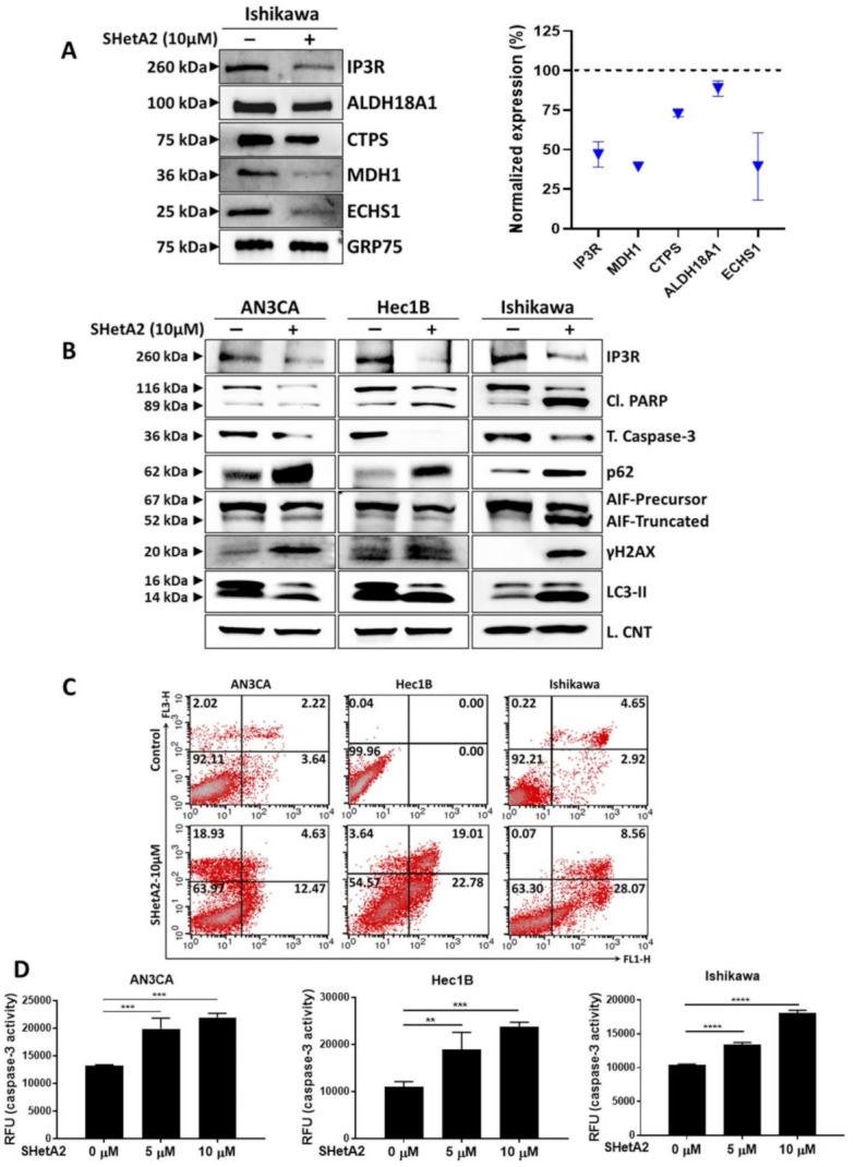
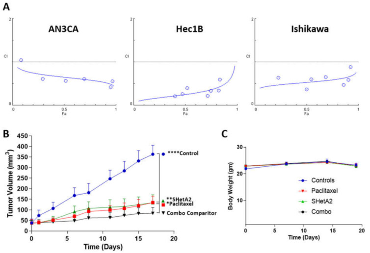
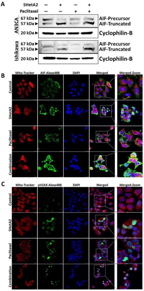
Endometrial cancer patients with advanced disease or high recurrence risk are treated with chemotherapy. Our objective was to evaluate the utility and mechanism of a novel drug, SHetA2, alone and in combination with paclitaxel, in endometrial cancer. SHetA2 targets the HSPA chaperone proteins, Grp78, hsc70, and mortalin, which have high mutation rates in endometrial cancer. SHetA2 effects on cancerous phenotypes, mitochondria, metabolism, protein expression, mortalin/client protein complexes, and cell death were evaluated in AN3CA, Hec13b, and Ishikawa endometrial cancer cell lines, and on growth of Ishikawa xenografts. In all three cell lines, SHetA2 inhibited anchorage-independent growth, migration, invasion, and ATP production, and induced G1 cell cycle arrest, mitochondrial damage, and caspase- and apoptosis inducing factor (AIF)-mediated apoptosis. These effects were associated with altered levels of proteins involved in cell cycle regulation, mitochondrial function, protein synthesis, endoplasmic reticulum stress, and metabolism; disruption of mortalin complexes with mitochondrial and metabolism proteins; and inhibition of oxidative phosphorylation and glycolysis. SHetA2 and paclitaxel exhibited synergistic combination indices in all cell lines and exerted greater xenograft tumor growth inhibition than either drug alone. SHetA2 is active against endometrial cancer cell lines in culture and in vivo and acts synergistically with paclitaxel.

Keywords: SHetA2; apoptosis inducing factor; cell cycle arrest; endometrial cancer; glycolysis; metabolism; mitochondria; oxidative phosphorylation; paclitaxel; xenograft.


Conflict of interest statement

The authors declare no conflict of interest.

Figures

Figure 1
Figure 1
SHetA2 attenuates cancerous phenotypes of endometrial cancer cell lines: (A) Soft agar colony formation assay of endometrial cancer cells treated with 5 µM SHetA2 or vehicle. After 3 weeks of culture, colonies were imaged (left panel) and counted (right panel) using q GelCount colony counter. Representative images scanned at 4× magnification are shown. The controls were set as 100% and data are shown as mean ± SD. (B) Wound healing scratch assay of endometrial cancer cells treated with 5 µM SHetA2 or vehicle. Images were captured at the indicated time points. (C) Invasion assays were conducted using the Matrigel-coated transwell chamber. Representative images of transwells showing invasion capacity of Hec1B and Ishikawa endometrial cancer cell in the presence of SHetA2 or vehicle are shown (left panel). The number of invaded cells were counted with a cell counter and shown (right panel). * p ≤ 0.05, ** p ≤ 0.01, *** p ≤ 0.001 when compared with respective controls.
Figure 2
Figure 2
SHetA2 induces cell cycle arrest and alters expression of cell cycle regulatory proteins: (A) Flow cytometry analysis of endometrial cancer cells treated with SHetA2 (10 µM) or vehicle for 24 h followed by PI staining. Representative images of cell cycle distributions are shown (upper panel). Percentages of the cell population at different cell cycle phases were calculated from three independent experiments and shown in the bar graph (Lower panel). (B) Western blot analysis of cell cycle regulatory proteins isolated from endometrial cancer cells treated with SHetA2 (10 µM) or vehicle for 24 h. * p ≤ 0.05, *** p ≤ 0.001 when compared with respective controls.
Figure 3
Figure 3
SHetA2 affects endometrial cancer cell mitochondria and metabolism: (A) JC-1 assay (ratio of aggregated JC-1 to monomer JC-1) in endometrial cancer cells treated with SHetA2 (10 µM) or vehicle. (B) Western blot analysis of mitochondrial dynamics-related proteins in endometrial cancer cells treated with or without SHetA2 (10 µM) for 24 h. (C) Metabolic viability of endometrial cancer cells treated with various doses of SHetA2 for 24, 48, and 72 h. The results are shown as % viability as compared to control. (D) ATP assay of endometrial cancer cells treated with SHetA2 (5 and 10 µM) for 24 h. (E) OCR and (F) Energy phenotype of Ishikawa cells treated with SHetA2 (10 µM) or vehicle for 4 h and measured by the Seahorse Cell Mito-Stress Test and the XFe96 Analyzer. (G) Extracellular acidification rate (ECAR) and (H) energy phenotype of Ishikawa cells treated with SHetA2 (10 µM) or vehicle for 4 h and measured with the Seahorse glycolytic rate assay and the XFe96 Analyzer. *** p ≤ 0.001, **** p ≤ 0.0001 when compared with respective control.
Figure 4
Figure 4
Molecular mechanism of SHetA2: (A) Ishikawa cells were treated with SHetA2 (10 µM) for 4 h and co-immunoprecipitation was performed with mortalin antibody (Grp75)-tagged beads. Western blots (left panel) and densitometry analyses (right panel) was performed for IP3R, ALDH18A1, CTPS, MDH1, and ECHS1 to confirm the reduction of client protein co-immunoprecipitation. (B) Western blot analysis for the protein expression of IP3R, Cl. PARP, total caspase-3, P62, AIF, DNA damage marker (γH2AX) and LC3-II proteins in endometrial cancer cells treated with, or without, SHetA2 (10 µM) for 24 h. (C) Cell apoptosis was detected by Annexin-V/PI combined labeling with flow cytometry in endometrial cancer cells 24 h after treatment with SHetA2 (10 µM) or vehicle. (D) Caspase-3 activity assay for endometrial cancer cells treated with indicated dose of SHetA2 for 24 h. ** p ≤ 0.01, *** p ≤ 0.001, **** p ≤ 0.0001 when compared with respective control.
Figure 5
Figure 5
SHetA2 enhances paclitaxel activity without toxicity: (A) CIs for endometrial cancer cell lines treated with a series of 2-fold dilutions of SHetA2 and paclitaxel in their 1:1 ratio of their IC50 concentrations in parallel with single drug treatments. After 72 h, the MTT assay was used to determine theFa’s, and CompuSyn was used to determine the CI’s. CI points below the dotted line indicate synergy. (B,C) Mice bearing Ishikawa-endometrial cancer xenografts were treated with oral 60 mg/kg/day SHetA2, 10 mg/kg/week i.p. paclitaxel, combination, or vehicle control. Average tumor volumes (B) and body weights (C) throughout the study are shown. ANOVA comparison of all treatment groups to the SHetA2 plus paclitaxel treated group (Combo comparator) * p < 0.05, ** p < 0.01 and **** p < 0.0001.
Figure 6
Figure 6
SHetA2 induces nuclear translocation of AIF, while both SHetA2 and paclitaxel induce DNA damage: (A) Western blot analysis for AIF expression in AN3CA and Ishikawa cells treated with SHetA2 or paclitaxel or their combination. (B,C) Representative immunofluorescence images for Mitotracker (red) and AIF (Green, B), or γH2AX (C) show their localization in AN3CA and Ishikawa cells treated with SHetA2 or paclitaxel or their combination. DAPI (Blue) staining was used as nuclear stain. Representative images taken at 63× magnification are shown. The left-most panels represent enlarged pictures of the square boxes shown in merged image.

Similar articles

Cited by

References

    1. Cancer Facts and Figures. [(accessed on 1 April 2021)]; Available online: https://www.cancer.org/research/cancer-facts-statistics/all-cancer-facts....
    1. Bray F., Ferlay J., Soerjomataram I., Siegel R.L., Torre L.A., Jemal A. Global cancer statistics 2018: GLOBOCAN estimates of incidence and mortality worldwide for 36 cancers in 185 countries. CA Cancer J. Clin. 2018;68:394–424. doi: 10.3322/caac.21492. - DOI - PubMed
    1. Aune D., Navarro Rosenblatt D.A., Chan D.S., Vingeliene S., Abar L., Vieira A.R., Greenwood D.C., Bandera E.V., Norat T. Anthropometric factors and endometrial cancer risk: A systematic review and dose-response meta-analysis of prospective studies. Ann. Oncol. 2015;26:1635–1648. doi: 10.1093/annonc/mdv142. - DOI - PubMed
    1. Arem H., Pfeiffer R.M., Moore S.C., Irwin M.L., LaMonte M.J., Sarto G.E., Nassir R., Luo J., Chlebowski R.T., Brinton L.A., et al. Post-diagnosis body mass index and mortality among women diagnosed with endometrial cancer: Results from the Women’s Health Initiative. PLoS ONE. 2017;12:e0171250. doi: 10.1371/journal.pone.0171250. - DOI - PMC - PubMed
    1. Amant F., Moerman P., Neven P., Timmerman D., Van Limbergen E., Vergote I. Endometrial cancer. Lancet. 2005;366:491–505. doi: 10.1016/S0140-6736(05)67063-8. - DOI - PubMed

LinkOut - more resources