

Anticancer Potential of Biogenic Silver Nanoparticles: A Mechanistic Study
- PMID: 34066092
- PMCID: PMC8151171
- DOI: 10.3390/pharmaceutics13050707
Anticancer Potential of Biogenic Silver Nanoparticles: A Mechanistic Study
Abstract
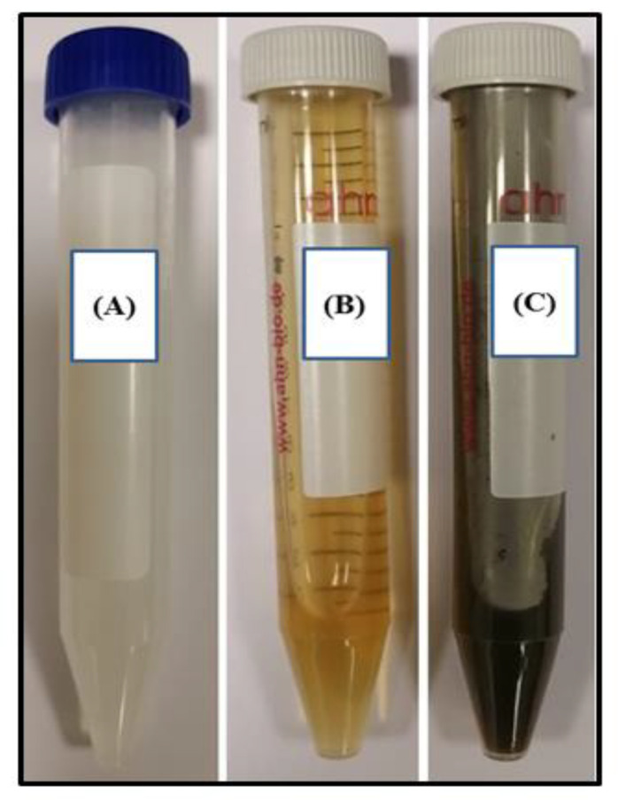
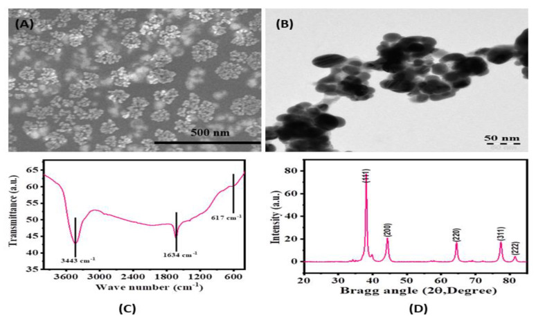
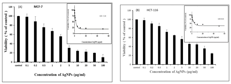
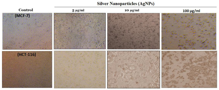
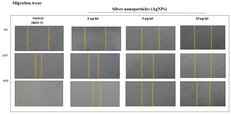
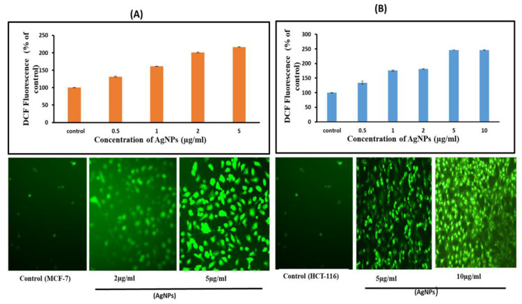
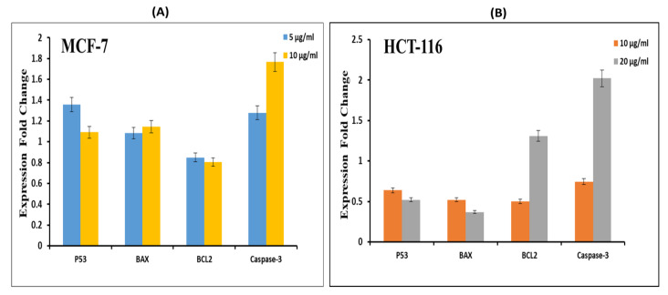
The continuous loss of human life due to the paucity of effective drugs against different forms of cancer demands a better/noble therapeutic approach. One possible way could be the use of nanostructures-based treatment methods. In the current piece of work, we have synthesized silver nanoparticles (AgNPs) using plant (Heliotropiumbacciferum) extract using AgNO3 as starting materials. The size, shape, and structure of synthesized AgNPs were confirmed by various spectroscopy and microscopic techniques. The average size of biosynthesized AgNPs was found to be in the range of 15 nm. The anticancer potential of these AgNPs was evaluated by a battery of tests such as MTT, scratch, and comet assays in breast (MCF-7) and colorectal (HCT-116) cancer models. The toxicity of AgNPs towards cancer cells was confirmed by the expression pattern of apoptotic (p53, Bax, caspase-3) and antiapoptotic (BCl-2) genes by RT-PCR. The cell viability assay showed an IC50 value of 5.44 and 9.54 µg/mL for AgNPs in MCF-7 and HCT-116 cell lines respectively. We also observed cell migration inhibiting potential of AgNPs in a concentration-dependent manner in MCF-7 cell lines. A tremendous rise (150-250%) in the production of ROS was observed as a result of AgNPs treatment compared with control. Moreover, the RT-PCR results indicated the difference in expression levels of pro/antiapoptotic proteins in both cancer cells. All these results indicate that cell death observed by us is mediated by ROS production, which might have altered the cellular redox status. Collectively, we report the antimetastasis potential of biogenic synthesized AgNPs against breast and colorectal cancers. The biogenic synthesis of AgNPs seems to be a promising anticancer therapy with greater efficacy against the studied cell lines.
Keywords: ROS; RT-PCR; cytotoxicity; scratch assay; silver nanoparticles.
Conflict of interest statement
The authors report no conflicts of interest in this work.
Figures

Similar articles
-
Silver Nanoparticles Phytofabricated through Azadirachta indica: Anticancer, Apoptotic, and Wound-Healing Properties.Antibiotics (Basel). 2023 Jan 9;12(1):121. doi: 10.3390/antibiotics12010121. Antibiotics (Basel). 2023. PMID: 36671322 Free PMC article.
-
Potential anticancer activity of biogenic silver nanoparticles using leaf extract of Rhynchosia suaveolens: an insight into the mechanism.Artif Cells Nanomed Biotechnol. 2018;46(sup1):104-114. doi: 10.1080/21691401.2017.1414824. Epub 2018 Jan 4. Artif Cells Nanomed Biotechnol. 2018. PMID: 29301413
-
A nanotechnology-based new approach in the treatment of breast cancer: Biosynthesized silver nanoparticles using Cuminum cyminum L. seed extract.J Photochem Photobiol B. 2020 Jul;208:111902. doi: 10.1016/j.jphotobiol.2020.111902. Epub 2020 May 20. J Photochem Photobiol B. 2020. PMID: 32470714
-
Biogenic silver nanoparticles synthesized using bracken fern inhibits cell proliferation in HCT-15 cells through induction of apoptosis pathway and overexpression of heat shock proteins.J Genet Eng Biotechnol. 2024 Dec;22(4):100428. doi: 10.1016/j.jgeb.2024.100428. Epub 2024 Oct 21. J Genet Eng Biotechnol. 2024. PMID: 39674645 Free PMC article.
-
A Bottom-Up Synthesis Approach to Silver Nanoparticles Induces Anti-Proliferative and Apoptotic Activities Against MCF-7, MCF-7/TAMR-1 and MCF-10A Human Breast Cell Lines.Molecules. 2020 Sep 22;25(18):4332. doi: 10.3390/molecules25184332. Molecules. 2020. PMID: 32971740 Free PMC article.
Cited by
-
Silver Nanoparticles Inhibit Metastasis of 4T1 Tumor in Mice after Intragastric but Not Intravenous Administration.Materials (Basel). 2022 May 27;15(11):3837. doi: 10.3390/ma15113837. Materials (Basel). 2022. PMID: 35683135 Free PMC article.
-
Nanotechnology-based delivery systems to overcome drug resistance in cancer.Med Rev (2021). 2024 Feb 20;4(1):5-30. doi: 10.1515/mr-2023-0058. eCollection 2024 Feb. Med Rev (2021). 2024. PMID: 38515777 Free PMC article. Review.
-
Nano Uncaria gambir as Chemopreventive Agent Against Breast Cancer.Int J Nanomedicine. 2023 Aug 3;18:4471-4484. doi: 10.2147/IJN.S403385. eCollection 2023. Int J Nanomedicine. 2023. PMID: 37555190 Free PMC article.
-
Evaluation of biogenically synthesized MgO NPs anticancer activity against breast cancer cells.Saudi J Biol Sci. 2024 Jan;31(1):103874. doi: 10.1016/j.sjbs.2023.103874. Epub 2023 Nov 11. Saudi J Biol Sci. 2024. PMID: 38090134 Free PMC article.
-
Anticancer, antioxidant and antibacterial potential of L-Glutaminase (Streptomyces roseolus strain ZKB1) capped silver and zinc oxide nanoparticles and its molecular characterization.Bioresour Bioprocess. 2025 Mar 23;12(1):23. doi: 10.1186/s40643-025-00857-w. Bioresour Bioprocess. 2025. PMID: 40121594 Free PMC article.
References
-
- Islam B., Khan M.S., Husain F., Rehman M.T., Alzughaibi T., Abuzenadah A.M., Urooj M., Kamal M.A., Tabrez S. mTOR targeted cancer chemoprevention by flavonoids. Curr. Med. Chem. 2020;28 in press. - PubMed
-
- Park Y.H., Hwang C., Kim Y., Lee Y., Jeong D., Cho M. Antimicrobial effects of silver nanoparticles. Nanomedicine. 2007;3:95–101. - PubMed
LinkOut - more resources
Full Text Sources
Research Materials
Miscellaneous
