. 2021 May 12;10(5):1183.
doi: 10.3390/cells10051183.

Minimalistic In Vitro Culture to Drive Human Naive B Cell Differentiation into Antibody-Secreting Cells

Affiliations

Minimalistic In Vitro Culture to Drive Human Naive B Cell Differentiation into Antibody-Secreting Cells

Peter-Paul A Unger et al. Cells. .

Abstract

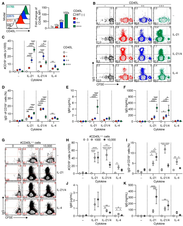
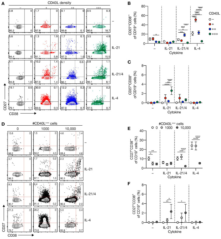
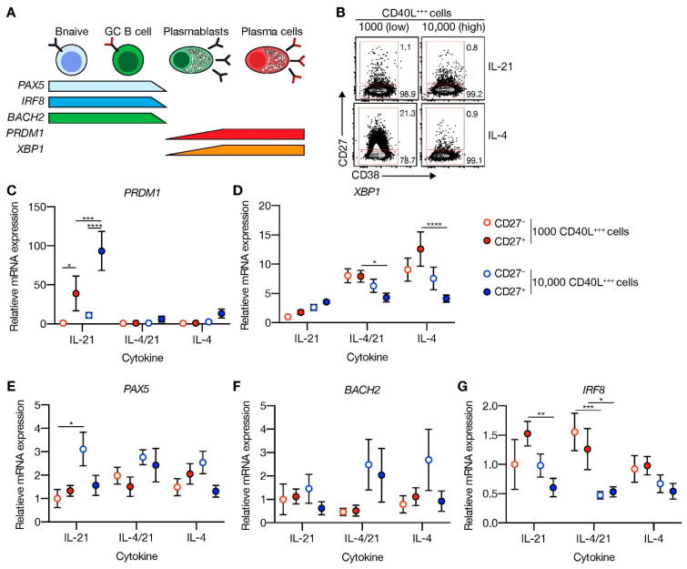
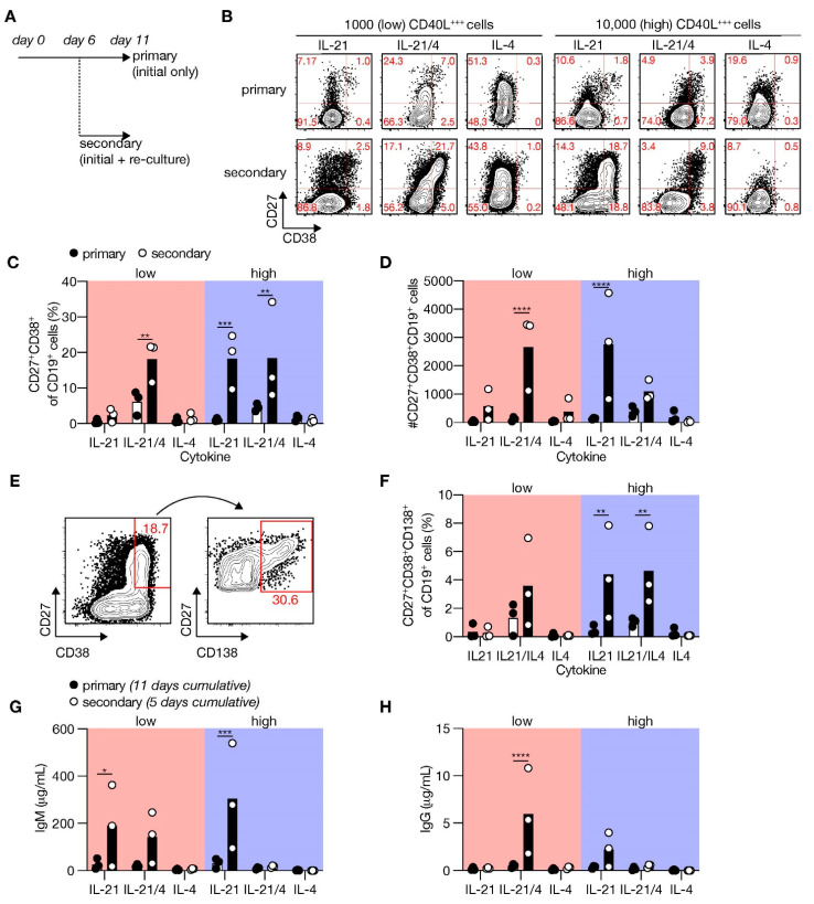
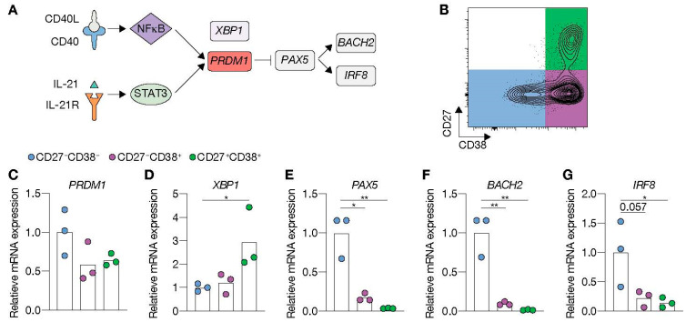
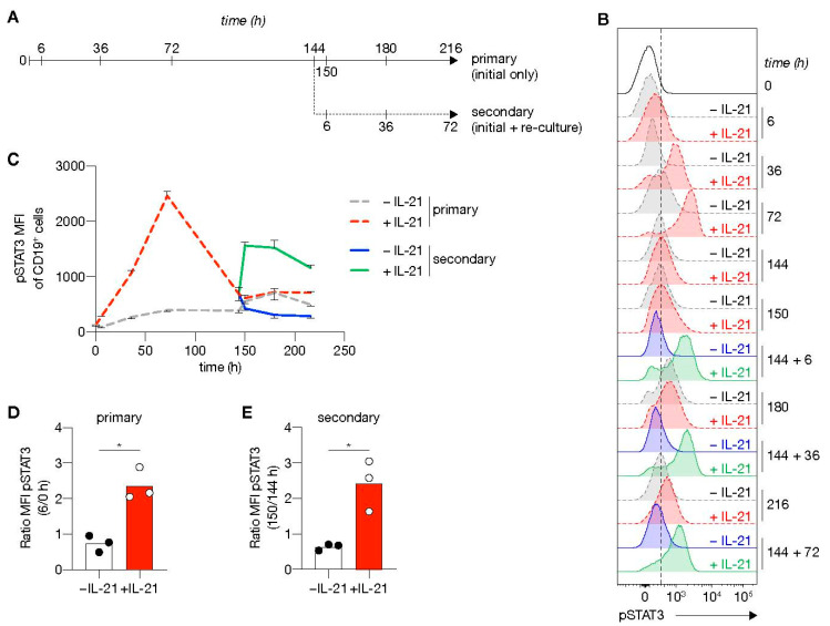
High-affinity antibody-secreting cells (ASC) arise from terminal differentiation of B-cells after coordinated interactions with T follicular helper (Tfh) cells in germinal centers (GC). Elucidation of cues promoting human naive B-cells to progress into ASCs is challenging, as this process is notoriously difficult to induce in vitro while maintaining enough cell numbers to investigate the differentiation route(s). Here, we describe a minimalistic in vitro culture system that supports efficient differentiation of human naive B-cells into antibody-secreting cells. Upon initial stimulations, the interplay between level of CD40 costimulation and the Tfh cell-associated cytokines IL-21 and IL-4 determined the magnitude of B-cell expansion, immunoglobulin class-switching and expression of ASC regulator PRDM1. In contrast, the B-cell-specific transcriptional program was maintained, and efficient ASC formation was hampered. Renewed CD40 costimulation and Tfh cytokines exposure induced rapid secondary STAT3 signaling and extensive ASC differentiation, accompanied by repression of B-cell identity factors PAX5, BACH2 and IRF8 and further induction of PRDM1. Our work shows that, like in vivo, renewed CD40L costimulation also induces efficient terminal ASC differentiation after initial B-cell expansion in vitro. This culture system for efficient differentiation of human naive B-cells into ASCs, while also maintaining high cell numbers, may form an important tool in dissecting human naive B-cell differentiation, thereby enabling identification of novel transcriptional regulators and biomarkers for desired and detrimental antibody formation in humans.

Keywords: cell differentiation; costimulatory molecules; cytokines; transcription factors.


Conflict of interest statement

The authors declare no conflict of interest. The funders had no role in the design of the study; in the collection, analyses or interpretation of data; in the writing of the manuscript; or in the decision to publish the results.

Figures

Figure 1
Figure 1
Level of CD40L expression and number of CD40L feeder cells supports human naive B-cell expansion and IgG class switching. (A) Representative histograms (left) of human CD40L expression stably transfected 3T3 fibroblasts expressing various amounts of human CD40L (+, ++ or +++) compared to non-transfected 3T3 cells (WT; −). Numbers indicate the mean fluorescence intensity (MFI) of CD40L expression. Fold changes compared to WT controls of CD40L MFI are shown in the right panel (data shown represent three separate experiments). (BD) Human CFSE-labeled naive B-cells were cultured on 3T3 cells expressing varying levels of CD40L with or without IL-21 and/or IL-4 for 6 days and analyzed for proliferation and class switching to surface IgG ((B); representative plots). The number of live CD19+ B-cells (C) and surface IgG-expression (D) was quantified after the cultures by flow cytometry analysis (n = 6). (EF) Cumulative secretion of IgM ((E); n = 9) and IgG ((F); n = 8) measured in culture supernatants after 11 days. (G) Representative plots of CFSE-labeled human naive B-cells cultured on 0/1000/10,000 CD40L+++-expressing 3T3 cells (supplemented to 10,000 with WT 3T3 cells) with or without IL-21 and/or IL-4 for 6 days. (HI) Number of live CD19+ events ((C); n = 9) and the frequency of IgG+ cells ((D); n = 7) after culture on 0/1000/10,000 CD40L+++-expressing 3T3 cells, and cytokines for 6 days. (JK) Cumulative IgM ((F); n = 7) and IgG ((G); n = 7) levels measured in culture supernatants after 11 days. Data are shown as mean ± SEM of independent experiments. Single experiments were conducted in triplicate. Data were analyzed by a two-way ANOVA followed by Tukey’s multiple comparison test. * P ≤ 0.05, ** P ≤ 0.01, *** P ≤ 0.001, **** P ≤ 0.0001.
Figure 2
Figure 2
Level of CD40L expression and number of CD40L feeder cells regulates differentiation of human naive B-cell into CD27+CD38 B-cells and CD27+CD38+ antibody-secreting cells. (A) Representative of FACS plot showing CD27 and CD38 expression among human naive B-cells cultured on CD40L-expressing 3T3 cells (as in Figure 1A) with or without IL-21 and/or IL-4 for 11 days. (B,C) The frequency of CD27+CD38 (B) and CD27+CD38+ (C) populations were analyzed 11 days after culture on CD40L-expressing 3T3 cells and cytokines. (D) Representative plots of human naive B-cells cultured on 0/1000/10,000 CD40L+++-expressing 3T3 cells (as in Figure 1A) supplemented to 10,000 with WT with or without IL-21 and/or IL-4 for 11 days. (E,F) The frequency of CD27+CD38 (E) and CD27+CD38+ (F) cells 11 days after culture on different numbers of CD40L+++-expressing 3T3 cells and cytokines. Data are shown as the mean ± SEM. (n = 6 (AC) and n = 7 (DF) independent experiments). Single experiments were conducted in triplicate. Data were analyzed by a two-way ANOVA followed by Tukey’s multiple comparison test. * P ≤ 0.05, ** P ≤ 0.01, *** P ≤ 0.001, **** P ≤ 0.0001.
Figure 3
Figure 3
CD40 costimulation together with IL-21 signaling induces PRDM1 expression and does not extinguish the B-cell lineage transcriptional program. (A) Schematic overview of the cellular stages and important transcription factors involved in B-cells differentiation from naive to antibody-secreting plasma cell (GC = germinal center). (B) Representative plots of human naive B-cells cultured on 1000 (low; supplemented with 9000 WT 3T3 cells) or 10,000 (high) CD40L+++-expressing 3T3 cells (as in Figure 1A) with or without IL-21 and/or IL-4 for 9 days. Subsequently, CD27CD38 (CD27) and CD27+CD38 (CD27+) cells were purified by cell sorting. (CG) Expression of PRMD1 (C), XBP1 (D), PAX5 (E), BACH2 (F) and IRF8 (G) mRNA in the sorted populations were analyzed by qPCR and related to levels present in the CD27 cells that were cultured on 1000 (low) CD40L+++-expressing 3T3 cells together with IL-21. Data are shown as mean ± SEM. (n = 5 independent experiments). Single experiments were conducted in triplicate. Data were analyzed by a two-way ANOVA followed by Tukey’s multiple comparison test. * P ≤ 0.05, ** P ≤ 0.01, *** P ≤ 0.001, **** P ≤ 0.0001.
Figure 4
Figure 4
Renewed CD40 costimulation and Tfh cytokines induce in vitro differentiation of human naive B-cells into antibody-secreting cells (ASCs). (A) Human B-cells cultured on 1000 (low; supplemented with 9000 WT 3T3 cells) or 10,000 (high) CD40L+++-expressing 3T3 cells (as in Figure 1A) with or without IL-21 and/or IL-4 for 11 days (primary—initial only stimulation). Alternatively, primary cultures were harvested after 6 days, and secondary cultures were initiated for 5 days with the same number of CD40L+++-expressing 3T3 cells and similar cytokine environments as in the primary culture. (B) Representative of FACS plots showing CD27 and CD38 expression among human B-cells cultured as in A. (C,D) The frequency (C) and number (D) of CD27+CD38+ cells were analyzed 11 days after initial only, or initial + secondary culture. (E) Representative plots of CD138 expression within the CD27+CD28+ ASC population. (F) The frequencies of CD138+ plasma cells were determined 11 days after initial only or initial + secondary culture. (G,H) Cumulative secretion of IgM (G) and IgG (H) measured in culture supernatants 11 days after initial only or 5 days after initial + secondary culture. Each data point represents the mean of an individual experiment (n = 3) with triplicate measurements. Mean values are represented as bars. p-values were calculated using multiple t-tests. * P ≤ 0.05, ** P ≤ 0.01, *** P ≤ 0.001, **** P ≤ 0.0001.
Figure 5
Figure 5
Renewed CD40 costimulation and IL-21 signaling drives antibody-secreting cell (ASC) differentiation and represses transcriptional program related to the B-cell fate. (A) Schematic representation of BLIMP1 activation upon CD40 and IL-21 receptor (IL-21R) signaling. STAT3, together with NFκB, regulates BLIMP1 expression. BLIMP1 downregulates PAX5 expression and, consequently, its downstream targets BACH2 and IRF8. (BG) Human naive B-cells were cultured on 10,000 (high) CD40L+++-expressing 3T3 cells (as in Figure 1A) for 6 days with IL-21. After 6 days, secondary cultures were initiated for 5 days with the initial stimuli. (B) Subsequently, CD27CD38, CD27CD38+ and CD27+CD38+ cell populations were sort purified. (CG) Expression of PRMD1 (C), XBP1 (D), PAX5 (E), BACH2 (F) and IRF8 (G) mRNA in sorted populations were analyzed by qPCR and related to levels present in CD27CD38 cells. Each data point represents the mean of an individual experiment (n = 3) with triplicate measurements. Mean values are represented as bars. p values were calculated using RM one-way ANOVA followed by Tukey’s multiple comparison test. * P ≤ 0.05, ** P ≤ 0.01.
Figure 6
Figure 6
STAT3 is rapidly re-phosphorylated upon re-stimulation using CD40 costimulation and IL-21 signaling. (A) Overview of the time points sampled from primary and secondary human B-cell cultures. Human naive B-cells were cultured on 10,000 CD40L+++-expressing 3T3 cells (as in Figure 1A) with or without IL-21 for 6 days. After 6 days, secondary cultures were initiated for 3 days with 10,000 CD40L+++-expressing 3T3 cells with(out) IL-21 (n = 3). (B,C) Representative plot of pSTAT3 levels (B) and quantification (C) at baseline and 6, 36, 72, 144, 150, 180 and 216 h in primary culture (dotted gray w/o IL-21 and red lines with IL-21); and 6, 36 and 72 in secondary culture (solid blue w/o IL-21 and green lines with IL-21). Data are shown as mean ± SEM (n = 3 independent experiments). Single experiments were conducted in triplicate. (D, E) Fold induction of pSTAT3 6 h in primary (D) and secondary (E) cultures relative to time point 0 or 144 h. Each data point represents the mean of an individual experiment (n = 3) with triplicate measurements. Mean values are represented as bars. p values were calculated using paired t-test. * P ≤ 0.05.

References

    1. Zimring J.C., Stowell S.R., Johnsen J.M., Hendrickson J.E. Effects of genetic, epigenetic, and environmental factors on alloimmunization to transfused antigens: Current paradigms and future considerations. Transfus. Clin. Biol. 2012;19:125–131. doi: 10.1016/j.tracli.2012.03.002. - DOI - PubMed
    1. Zimring J.C., Hudson K.E. Cellular immune responses in red blood cell alloimmunization. Hematology. 2016;2016:452–456. doi: 10.1182/asheducation-2016.1.452. - DOI - PMC - PubMed
    1. Laffitte E., Skaria M., Jaunin F., Tamm K., Saurat J.-H., Favre B., Borradori L. Autoantibodies to the extracellular and intracellular domain of bullous pemphigoid 180, the putative key autoantigen in bullous pemphigoid, belong predominantly to the IgG1 and IgG4 subclasses. Br. J. Dermatol. 2001;144:760–768. doi: 10.1046/j.1365-2133.2001.04130.x. - DOI - PubMed
    1. Gilhus N.E., Skeie G.O., Romi F., Lazaridis K., Zisimopoulou P., Tzartos S. Myasthenia gravis—Autoantibody characteristics and their implications for therapy. Nat. Rev. Neurol. 2016;12:259–268. doi: 10.1038/nrneurol.2016.44. - DOI - PubMed
    1. Berentsen S., Sundic T. Red Blood Cell Destruction in Autoimmune Hemolytic Anemia: Role of Complement and Potential New Targets for Therapy. BioMed Res. Int. 2015;2015:363278. doi: 10.1155/2015/363278. - DOI - PMC - PubMed

Publication types

MeSH terms