Recurrence in Oral Premalignancy: Clinicopathologic and Immunohistochemical Analysis
- PMID: 34066207
- PMCID: PMC8151734
- DOI: 10.3390/diagnostics11050872
Recurrence in Oral Premalignancy: Clinicopathologic and Immunohistochemical Analysis
Abstract
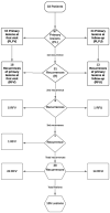
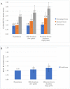
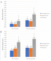
Oral leukoplakia (OL) has a propensity for recurrence and malignant transformation (MT). Herein, we evaluate sociodemographic, clinical, microscopic and immunohistochemical parameters as predictive factors for OL recurrence, also comparing primary lesions (PLs) with recurrences. Thirty-three patients with OL, completely removed either by excisional biopsy or by laser ablation following incisional biopsy, were studied. Selected molecules associated with the STAT3 oncogenic pathway, including pSTAT3, Bcl-xL, survivin, cyclin D1 and Ki-67, were further analyzed. A total of 135 OL lesions, including 97 PLs and 38 recurrences, were included. Out of 97 PLs, 31 recurred at least once and none of them underwent MT, during a mean follow-up time of 48.3 months. There was no statistically significant difference among the various parameters in recurrent vs. non-recurrent PLs, although recurrence was most frequent in non-homogeneous lesions (p = 0.087) and dysplastic lesions recurred at a higher percentage compared to hyperplastic lesions (34.5% vs. 15.4%). Lower levels of Bcl-xL and survivin were identified as significant risk factors for OL recurrence. Recurrences, although smaller and more frequently homogeneous and non-dysplastic compared to their corresponding PLs, exhibited increased immunohistochemical expression of oncogenic molecules, especially pSTAT3 and Bcl-xL. Our results suggest that parameters associated with recurrence may differ from those that affect the risk of progression to malignancy and support OL management protocols favoring excision and close monitoring of all lesions.
Keywords: Bcl-xL; Ki-67; STAT3; cyclin D1; laser ablation; oral leukoplakia; oral potentially malignant disorders; predictive biomarkers; recurrence; survivin.
Conflict of interest statement
The authors declare no conflict of interest.
Figures


Similar articles
-
Recurrence rates after surgical removal of oral leukoplakia-A prospective longitudinal multi-centre study.PLoS One. 2019 Dec 6;14(12):e0225682. doi: 10.1371/journal.pone.0225682. eCollection 2019. PLoS One. 2019. PMID: 31810078 Free PMC article.
-
Expression of phosphorylated Stat3, cyclin D1 and Bcl-xL in extramammary Paget disease.Br J Dermatol. 2006 May;154(5):926-32. doi: 10.1111/j.1365-2133.2005.06951.x. Br J Dermatol. 2006. PMID: 16634897
-
Oral leukoplakia manifests differently in smokers and non-smokers.Braz Oral Res. 2012 Nov-Dec;26(6):543-9. doi: 10.1590/s1806-83242012005000024. Epub 2012 Sep 27. Braz Oral Res. 2012. PMID: 23019086
-
STAT3 mRNA and protein expression in colorectal cancer: effects on STAT3-inducible targets linked to cell survival and proliferation.J Clin Pathol. 2007 Feb;60(2):173-9. doi: 10.1136/jcp.2005.035113. J Clin Pathol. 2007. PMID: 17264243 Free PMC article.
-
Laser surgery as a treatment for oral leukoplakia.Oral Oncol. 2003 Dec;39(8):759-69. doi: 10.1016/s1368-8375(03)00043-5. Oral Oncol. 2003. PMID: 13679199 Review.
Cited by
-
Prevalence and Genotyping of HPV in Oral Squamous Cell Carcinoma in Northern Brazil.Pathogens. 2022 Sep 27;11(10):1106. doi: 10.3390/pathogens11101106. Pathogens. 2022. PMID: 36297163 Free PMC article.
-
Photodynamic therapy (PDT) for oral leukoplakia: a systematic review and meta-analysis of single-arm studies examining efficacy and subgroup analyses.BMC Oral Health. 2023 Aug 13;23(1):568. doi: 10.1186/s12903-023-03294-3. BMC Oral Health. 2023. PMID: 37574560 Free PMC article.
-
Podoplanin Expression Independently and Jointly with Oral Epithelial Dysplasia Grade Acts as a Potential Biomarker of Malignant Transformation in Oral Leukoplakia.Biomolecules. 2022 Apr 19;12(5):606. doi: 10.3390/biom12050606. Biomolecules. 2022. PMID: 35625534 Free PMC article.
References
-
- Warnakulasuriya S., Kujan O., Aguirre-Urizar J.M., Bagan J.V., González-Moles M.Á., Kerr A.R., Lodi G., Mello F.W., Monteiro L., Ogden G.R., et al. Oral potentially malignant disorders: A consensus report from an international seminar on nomenclature and classification, convened by the WHO Collaborating Centre for Oral Cancer. Oral Dis. 2020 doi: 10.1111/odi.13704. - DOI - PubMed
LinkOut - more resources
Full Text Sources
Research Materials
Miscellaneous

