Review
. 2021 May 8;22(9):4994.
doi: 10.3390/ijms22094994.

Neurons and Glia Interplay in α-Synucleinopathies

Affiliations
Review

Neurons and Glia Interplay in α-Synucleinopathies

Panagiota Mavroeidi et al. Int J Mol Sci. .

Abstract

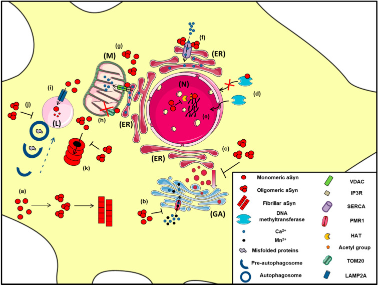
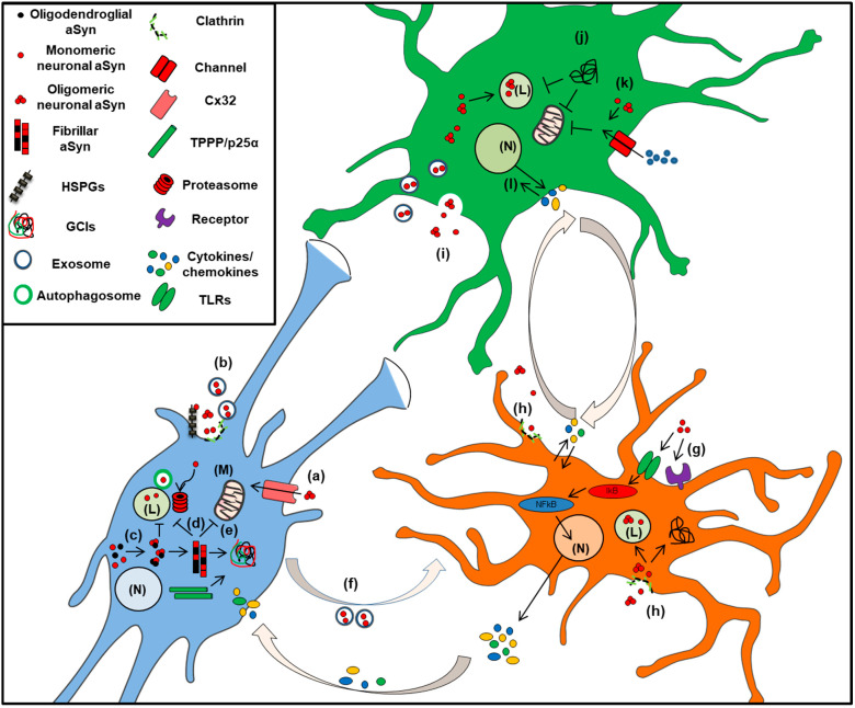
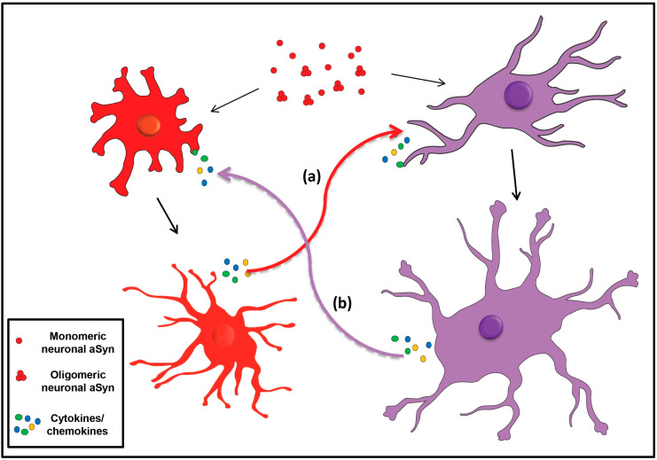
Accumulation of the neuronal presynaptic protein alpha-synuclein within proteinaceous inclusions represents the key histophathological hallmark of a spectrum of neurodegenerative disorders, referred to by the umbrella term a-synucleinopathies. Even though alpha-synuclein is expressed predominantly in neurons, pathological aggregates of the protein are also found in the glial cells of the brain. In Parkinson's disease and dementia with Lewy bodies, alpha-synuclein accumulates mainly in neurons forming the Lewy bodies and Lewy neurites, whereas in multiple system atrophy, the protein aggregates mostly in the glial cytoplasmic inclusions within oligodendrocytes. In addition, astrogliosis and microgliosis are found in the synucleinopathy brains, whereas both astrocytes and microglia internalize alpha-synuclein and contribute to the spread of pathology. The mechanisms underlying the pathological accumulation of alpha-synuclein in glial cells that under physiological conditions express low to non-detectable levels of the protein are an area of intense research. Undoubtedly, the presence of aggregated alpha-synuclein can disrupt glial function in general and can contribute to neurodegeneration through numerous pathways. Herein, we summarize the current knowledge on the role of alpha-synuclein in both neurons and glia, highlighting the contribution of the neuron-glia connectome in the disease initiation and progression, which may represent potential therapeutic target for a-synucleinopathies.

Keywords: a-Synuclein; aggregation; astrocytes; inclusions; microglia; neurons; oligodendroglia; seeding.


Conflict of interest statement

The authors declare no conflict of interest.

Figures

Figure 1
Figure 1
The role of aSyn at the presynaptic terminal. A schematic representation depicting of aSyn physiological and pathological effects at the synapse: (a) aSyn reduces the activity of tyrosine hydroxylase (TH), the enzyme responsible for catalyzing the conversion of L-Tyrosine to L-DOPA, thus impairing dopamine biosynthesis, (b) Increased levels of aSyn inhibit VMAT2, which is responsible for the uptake of monoamines (such as dopamine) into synaptic vesicles, (c) aSyn associates with synaptic vesicle membranes and regulates the SNARE-dependent vesicle fusion and neurotransmitter release, (d) Soluble aSyn interacts with the dopaminergic transporter DAT and decreases its amount on the plasma membrane, thus regulating the dopamine re-uptake from the synapse. However, aSyn aggregates trigger DAT recruitment to the plasma membrane that leads to massive entry of dopamine, (e) aSyn aggregates interact with Na+/K+-ATPase (NKA) preventing the effective pump out of Na+ ions, (f) aSyn is secreted from neuronal cells partly via associating with exosomes, (g) Extracellular aSyn interacts with neuronal receptors (i.e., LAG3) for its internalization in neurons or (h) it is up-taken via endocytosis, (i) PD-linked A30P and A53T mutant aSyn form large membrane pores through which most cations (i.e., Ca2+) can pass non-selectively, (j) Extracellular aSyn activates the voltage-gated Ca2+ channels (VGCCs), resulting in increased Ca2+ influx, (k) Monomeric aSyn enters neuronal cells via passive diffusion or direct penetration of their plasma membrane.
Figure 2
Figure 2
The proposed intracellular effects of various aSyn conformations in neurons. A schematic representation of the aberrant interactions between the various aSyn species with intracellular organelles: (a) In the cytoplasm of neurons, aSyn monomers form oligomers that can eventually become fibrils, (b) Both unfolded and aggregated aSyn impair the function of PMR1, a Ca2+-transporting ATPase pump that regulates Ca2+ and Mn+2 levels in the Golgi apparatus (GA), resulting in elevated cytosolic Ca2+ levels, (c) Both WT and mutant A53T aSyn disrupt the vesicular transport from Endoplasmic Reticulum (ER) to Golgi (GA), (d) WT aSyn inhibits the transportation of methyltransferases from the cytoplasm to the nucleus (N), thus altering DNA methylation of the SNCA gene, (e) Inside the nucleus (N), aSyn inhibits histone acetylation via its direct binding to histones or by preventing the action of histone acetyltransferase (HAT) enzymes, thus interfering in the process of gene transcription, (f) In the ER, aSyn aggregates activate the Ca2+-ATPase SERCA, resulting in dysregulated Ca2+ homeostasis, (g) Both monomeric and oligomeric aSyn interact with Voltage-dependent anion channel 1 (VDAC1) and inositol triphosphate receptors (IP3Rs), the protein components involved in mitochondrial-associated ER membrane (MAM) and regulates the transmission of Ca2+ signals from the ER to mitochondria (M), (h) aSyn binds to TOM20, a mitochondrial import receptor subunit and inhibits normal protein import, (i) Normally, monomeric or dimeric forms of aSyn are degraded in the lysosome (L) via Chaperone Mediated Autophagy (CMA), following their interaction with LAMP2A. However, under pathological conditions, impairment of CMA has been proposed to lead to aSyn accumulation and subsequent cell toxicity, (j) Oligomeric aSyn and various misfolded proteins are cleared via macroautophagy, following the fusion of autophagosomes with the lysosome. Pathological aSyn has been shown to inhibit autophagosome maturation or their fusion with lysosomes, thus impairing autophagic flux, (k) Monomeric and oligomeric aSyn are degraded via the proteasome; however, under pathological conditions, increased levels of aSyn or even soluble aSyn oligomers may inhibit proteasomal function, leading to aSyn accumulation and the formation of insoluble aggregates.
Figure 3
Figure 3
A complicated neuron-glial interrelationship. (a) Oligomeric aSyn enters oligodendrocytes (blue) via binding to the gap junction protein connexin-32 (Cx32), (b) Neuronally-derived aSyn enters oligodendrocytes via clathrin-mediated endocytosis, exosomal transportation, or via binding to Heparan Sulfate ProteoGlycans (HSPGs), (c) Inside oligodendrocytes, neuronal aSyn (red) initiates the seeding of the endogenous oligodendroglial aSyn (black) and together with the oligodendroglial-specific TPPP/p25α protein, they lead to the formation of GCIs, (d) aSyn aggregates impair the proteolytic machineries of oligodendrocytes [(proteasome and lysosome (L)], (e) Misfolded aSyn leads to mitochondrial (M) dysfunction and subsequent cell toxicity in MSA, (f) Oligodendroglial-derived exosomes containing aSyn have been shown to transfer to microglial cells (orange), (g) Extracellular aSyn stimulates TLRs and other receptors (i.e., FcγR, P2X7 etc) to activate microglial transcription factors, such as NFkB, for the production of various pro-inflammatory cytokines (IL1β, TNFa) and chemokines that induce astrocyte (green) reactivity and oligodendroglial damage, (h) Neuronally-secreted aSyn is taken up by microglial cells via clathrin-mediated endocytosis and is then driven to the lysosome (L) for degradation. In pathological conditions, though, it accumulates into aSyn insoluble aggregates, (i) Free or exosome-associated aSyn released by neurons is transmitted to astrocytes via endocytosis and enters the lysosome (L) for its clearance, (j) In disease, aSyn aggregates are formed that lead to lysosomal (L) impairment and mitochondria (M) dysfunction, (k) In astrocytes, aSyn triggers the opening of channels (i.e., Cx43 and Panx1), leading to dysregulation in Ca2+ homeostasis and altered mitochondrial morphology, (l) Microglia-secreted cytokines and chemokines activate astrocytes to further produce pro-inflammatory signaling molecules and enhance neurotoxicity.
Figure 4
Figure 4
Crosstalk between astrocytes and microglia in a-synucleinopathies. Monomeric or oligomeric aSyn released by neurons is taken-up by astrocytes (purple) and microglia (red), which are then activated and secrete various cytokines or chemokines. (a) According to the prevailing hypothesis, the released pro-inflammatory molecules by microglia trigger the activation of astrocytes (red arrow), leading to excessive inflammation and neurotoxicity. (b) Conversely, aSyn can directly activate astrocytes to secrete pro-inflammatory cytokines or chemokines that recruit and activate microglia (purple arrow) resulting in excessive neuroinflammation.

Similar articles

Cited by

References

    1. Uversky V.N. A protein-chameleon: Conformational plasticity of alpha-synuclein, a disordered protein involved in neurodegenerative disorders. J. Biomol. Struct. Dyn. 2003;21:211–234. doi: 10.1080/07391102.2003.10506918. - DOI - PubMed
    1. Spillantini M.G., Crowther R.A., Jakes R., Hasegawa M., Goedert M. alpha-Synuclein in filamentous inclusions of Lewy bodies from Parkinson’s disease and dementia with lewy bodies. Proc. Natl. Acad. Sci. USA. 1998;95:6469–6473. doi: 10.1073/pnas.95.11.6469. - DOI - PMC - PubMed
    1. Polymeropoulos M.H., Lavedan C., Leroy E., Ide S.E., Dehejia A., Dutra A., Pike B., Root H., Rubenstein J., Boyer R., et al. Mutation in the alpha-synuclein gene identified in families with Parkinson’s disease. Science. 1997;276:2045–2047. doi: 10.1126/science.276.5321.2045. - DOI - PubMed
    1. Chartier-Harlin M.C., Kachergus J., Roumier C., Mouroux V., Douay X., Lincoln S., Levecque C., Larvor L., Andrieux J., Hulihan M., et al. Alpha-synuclein locus duplication as a cause of familial Parkinson’s disease. Lancet. 2004;364:1167–1169. doi: 10.1016/S0140-6736(04)17103-1. - DOI - PubMed
    1. Kruger R., Kuhn W., Muller T., Woitalla D., Graeber M., Kosel S., Przuntek H., Epplen J.T., Schols L., Riess O. Ala30Pro mutation in the gene encoding alpha-synuclein in Parkinson’s disease. Nat. Genet. 1998;18:106–108. doi: 10.1038/ng0298-106. - DOI - PubMed

Substances

LinkOut - more resources