Review
. 2021 May 8;13(5):862.
doi: 10.3390/v13050862.

Hepatitis B Virus Pre-S Gene Deletions and Pre-S Deleted Proteins: Clinical and Molecular Implications in Hepatocellular Carcinoma

Affiliations
Review

Hepatitis B Virus Pre-S Gene Deletions and Pre-S Deleted Proteins: Clinical and Molecular Implications in Hepatocellular Carcinoma

Yueh-Te Lin et al. Viruses. .

Abstract

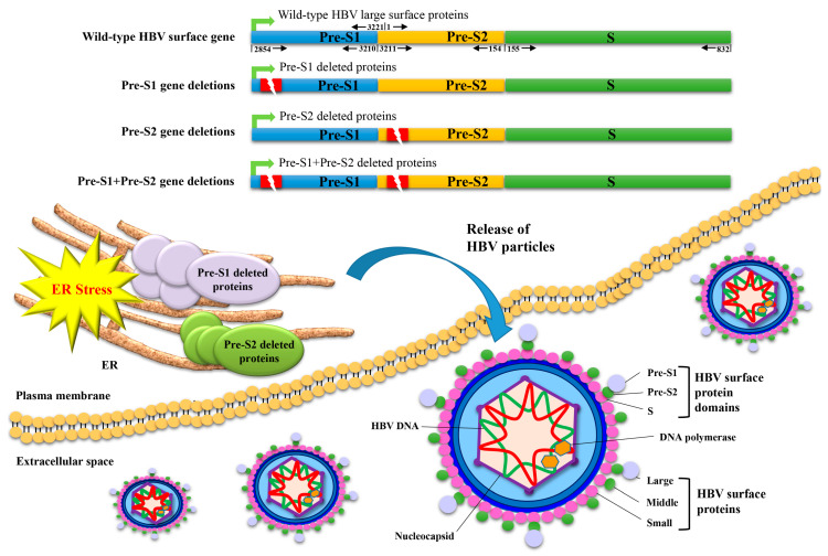
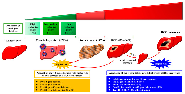
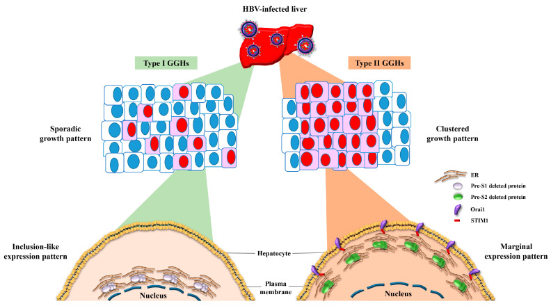
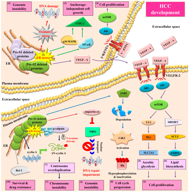
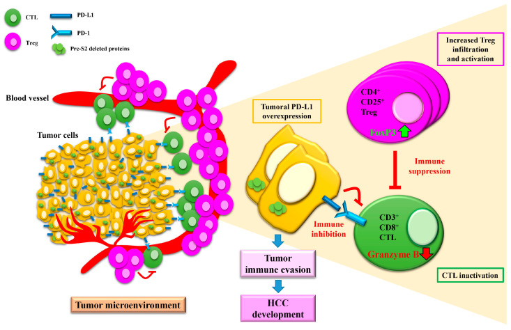
Hepatocellular carcinoma (HCC) is one of the most frequent and fatal human cancers worldwide and its development and prognosis are intimately associated with chronic infection with hepatitis B virus (HBV). The identification of genetic mutations and molecular mechanisms that mediate HBV-induced tumorigenesis therefore holds promise for the development of potential biomarkers and targets for HCC prevention and therapy. The presence of HBV pre-S gene deletions in the blood and the expression of pre-S deleted proteins in the liver tissues of patients with chronic hepatitis B and HBV-related HCC have emerged as valuable biomarkers for higher incidence rates of HCC development and a higher risk of HCC recurrence after curative surgical resection, respectively. Moreover, pre-S deleted proteins are regarded as important oncoproteins that activate multiple signaling pathways to induce DNA damage and promote growth and proliferation in hepatocytes, leading to HCC development. The signaling molecules dysregulated by pre-S deleted proteins have also been validated as potential targets for the prevention of HCC development. In this review, we summarize the clinical and molecular implications of HBV pre-S gene deletions and pre-S deleted proteins in HCC development and recurrence and highlight their potential applications in HCC prevention and therapy.

Keywords: biomarkers; hepatitis B virus; hepatocellular carcinoma; pre-S deleted proteins; pre-S gene deletions; targets.


Conflict of interest statement

The authors declare no conflict of interest.

Figures

Figure 1
Figure 1
Schematic representation of HBV pre-S gene deletions and the expression and ER retention of pre-S deleted proteins. The HBV surface gene consists of three gene segments (pre-S1, pre-S2, and S) and expresses three different sizes of surface proteins (small, middle, and large) that are, respectively, composed of domains encoded from the S segment, the pre-S2 and S segments, and all three gene segments, collectively constituting the envelope proteins of viral particles. The numbers shown above and below the wild-type surface gene indicate the nt positions of each intact gene segment in the circular HBV DNA genome, which starts at nt 1 and ends at nt 3221 in a clockwise direction. Deletion mutations (indicated as red rectangle boxes with a lightning symbol) that occur solely in the pre-S1 or pre-S2 gene segment lead to the expression of pre-S1 or pre-S2 deletion mutants of large surface proteins, respectively. Unlike wild-type large surface proteins that are released as constituents of the envelope proteins of HBV particles from host cells, both pre-S1 and pre-S2 deleted proteins are mainly retained in the host cell organelle ER and induce ER stress. In addition, deletion mutations that occur concurrently in the pre-S1 and pre-S2 gene segments (the so-called pre-S1 + pre-S2 gene deletions) may lead to the expression of large surface proteins harboring both pre-S1 and pre-S2 deletions (the so-called pre-S1 + pre-S2 deleted proteins), which may act in a manner like pre-S1 and pre-S2 deleted proteins—although the exact molecular function remains to be clarified. The HBV DNA polymerase associated with the viral DNA genome and the nucleocapsid encompassing the viral DNA genome are also shown in the diagram. Abbreviations: HBV, hepatitis B virus; ER, endoplasmic reticulum.
Figure 2
Figure 2
Schematic summary of the clinical association of HBV pre-S gene deletions with liver disease progression and HCC development and recurrence. In patients with chronic HBV infection, the prevalence of overall pre-S gene deletions (including pre-S1, pre-S2, and pre-S1 + pre-S2 gene deletions) in the blood gradually increases with liver disease progression from the high to intermediate to low replicative phases of chronic hepatitis B, to liver cirrhosis, and reaches the highest level at the stage of HCC development. The presence of pre-S gene deletions (pre-S1, pre-S2, and/or pre-S1 + pre-S2 gene deletions) or pre-S2 gene deletions between nts 38 and 55 (nt 38 to 55) predicts higher incidence of liver cirrhosis and HCC development. Moreover, in patients with HBV-related HCC, the presence of deletions spanning the pre-S2 gene segment, pre-S2 gene deletion at nts 1 to 54 (nt 1 to 54), pre-S2 gene deletions at ≥5%, or pre-S2 plus pre-S1+pre-S2 gene deletions at >25% in the blood or type II GGHs comprising ≥10% of hepatocytes in the liver tissues predicts the higher risk of HCC recurrence after curative surgical resection. Abbreviations: HCC, hepatocellular carcinoma; GGHs, ground glass hepatocytes.
Figure 3
Figure 3
Schematic representation of the growth patterns of GGHs and the expression patterns of HBV pre-S deleted proteins. In the liver tissues of patients with chronic HBV infection, two distinct types of GGHs (type I and type II) are identified as HBV-infected hepatocytes that express pre-S1 and pre-S2 deleted proteins, respectively. Type I GGHs display a scattered or sporadic growth pattern and express pre-S1 deleted proteins in a globular or inclusion-like pattern due to the accumulation of pre-S1 deleted proteins in the ER, whereas type II GGHs display a clustered growth pattern and express pre-S2 deleted proteins in a marginal pattern due to the accumulation of pre-S2 deleted proteins in the ER followed by STIM1-and Orai1-mediated connection of the ER and plasma membrane. Abbreviations: HBV, hepatitis B virus; GGHs, ground glass hepatocytes; ER, endoplasmic reticulum; STIM1, stromal interaction molecule 1; Orai1, calcium release-activated calcium modulator 1.
Figure 4
Figure 4
Schematic summary of signaling pathways activated by HBV pre-S deleted proteins and their implications in HCC development. In chronic HBV infection, both pre-S1 and pre-S2 deleted proteins accumulate in the ER and induce ER stress in hepatocytes (as highlighted by (1)). Through the mediation of ER stress, both types of pre-S deleted proteins can initiate three oncogenic signaling pathways, one involving oxidative stress to induce DNA damage and genomic instability (as highlighted by (2)), another involving NF-κB/p38 MAPK/COX-2 to enhance anchorage-independent cell growth (as highlighted by (3)), and the other involving VEGF-A/VEGFR-2/Akt/mTOR to promote cell proliferation (as highlighted by (4)). Through the mediation of mTOR activation, pre-S deleted proteins can further initiate two metabolism-related signaling pathways; one involving YY1/Myc/SLC2A1 to stimulate aerobic glycolysis (as highlighted by (5)) and the other involving SREBF1/ACLY/FADS2 to increase lipid biosynthesis (as highlighted by (6)), collectively contributing to cell proliferation. Furthermore, pre-S2 deleted proteins can additionally initiate four ER stress-dependent or -independent signaling pathways: one involving Ca2+/μ-calpain/ΔN-cyclin A to induce centrosome overduplication and chromosome instability (as highlighted by (7)), one involving JAB1/p27/Cdk2/Rb to promote cell cycle progression (as highlighted by (8)), one involving importin α1/NBS1 to inhibit DNA repair (as highlighted by (9)), and the other involving Bcl-2 to enhance cell survival and drug resistance (as highlighted by (10)). The combined effects of pre-S deleted proteins on DNA damage and genomic instability, anchorage-independent growth and proliferation, aerobic glycolysis and lipid biosynthesis, centrosome overduplication and chromosome instability, cell cycle progression, and cell survival result in HCC development. Abbreviations: HBV, hepatitis B virus; ER, endoplasmic reticulum; NF-κB, nuclear factor-κB; p38 MAPK, p38 mitogen-activated protein kinase; COX-2, cyclooxygenase-2; VEGF-A, vascular endothelial growth factor-A; VEGFR-2, vascular endothelial growth factor receptor-2; Akt, protein kinase B; mTOR, mammalian target of rapamycin; YY1, Yin Yang 1; SLC2A1, solute carrier family 2 (facilitated glucose transporter) member 1; SREBF1, sterol regulatory element-binding transcription factor 1; ACLY, adenosine triphosphate citrate lyase; FADS2, fatty acid desaturase 2; Ca2+, calcium; ΔN-cyclin A, N-terminus truncated cyclin A; JAB1, Jun activation domain-binding protein 1; Cdk2, cyclin-dependent kinase 2; Rb, retinoblastoma protein; NBS1, Nijmegen breakage syndrome 1; Bcl-2, B cell lymphoma-2; HCC, hepatocellular carcinoma.
Figure 5
Figure 5
Schematic representation of the hypothetical model of HBV pre-S2 deleted proteins in the regulation of the tumor immune microenvironment in HCC. In the tumor microenvironment of HCC patients positive for pre-S2 deleted proteins, the expression of PD-L1 in tumor cells is elevated, the infiltration and activity of CD4+CD25+ Tregs are increased (as shown by increased cell number and forkhead box P3 (Foxp3) expression), and the activity of CD3+CD8+ CTLs is decreased (as shown by decreased granzyme B expression and secretion). Tregs have been shown to exert “pro-tumor” activity by suppressing the activity of “anti-tumor” CTLs [57]. In addition, PD-L1 expressed in tumor cells has been shown to interact with its receptor, PD-1, expressed in CTLs, to dampen the attack of tumor cells by CTLs [58]. The combined effects of tumoral PD-L1 overexpression, increased Treg infiltration and activation, and CTL inactivation favor tumor cell evasion from host immune surveillance, contributing to HCC development in patients with pre-S2 deleted proteins. Abbreviations: Treg, regulatory T cell; Foxp3, forkhead box P3; CTL, cytotoxic T cell; PD-L1, programmed death ligand 1; PD-1, programmed death 1; HCC, hepatocellular carcinoma.

Similar articles

Cited by

References

    1. Llovet J.M., Zucman-Rossi J., Pikarsky E., Sangro B., Schwartz M., Sherman M., Gores G. Hepatocellular carcinoma. Nat. Rev. Dis. Primers. 2016;2:16018. doi: 10.1038/nrdp.2016.18. - DOI - PubMed
    1. Cheng K.-C., Lin W.-Y., Liu C.-S., Lin C.-C., Lai H.-C., Lai S.-W. Association of different types of liver disease with demographic and clinical factors. Biomedicine. 2016;6:1–7. doi: 10.7603/s40681-016-0016-2. - DOI - PMC - PubMed
    1. Kulik L., El-Serag H.B. Epidemiology and management of hepatocellular carcinoma. Gastroenterology. 2019;156:477–491. doi: 10.1053/j.gastro.2018.08.065. - DOI - PMC - PubMed
    1. Sung H., Ferlay J., Siegel R.L., Laversanne M., Soerjomataram I., Jemal A., Bray F. Global cancer statistics 2020: GLOBOCAN estimates of incidence and mortality worldwide for 36 cancers in 185 countries. CA Cancer J. Clin. 2021;68:394–424. doi: 10.3322/caac.21660. - DOI - PubMed
    1. Zhou L., Wang S.-B., Chen S.-G., Qu Q., Rui J.-A. Risk factors of recurrence and poor survival in curatively resected hepatocellular carcinoma with microvascular invasion. Adv. Clin. Exp. Med. 2020;29:887–892. doi: 10.17219/acem/76750. - DOI - PubMed

Publication types

MeSH terms