. 2021 May 7;22(9):4961.
doi: 10.3390/ijms22094961.

Methionine Diet Evoked Hyperhomocysteinemia Causes Hippocampal Alterations, Metabolomics Plasma Changes and Behavioral Pattern in Wild Type Rats

Affiliations

Methionine Diet Evoked Hyperhomocysteinemia Causes Hippocampal Alterations, Metabolomics Plasma Changes and Behavioral Pattern in Wild Type Rats

Maria Kovalska et al. Int J Mol Sci. .

Abstract

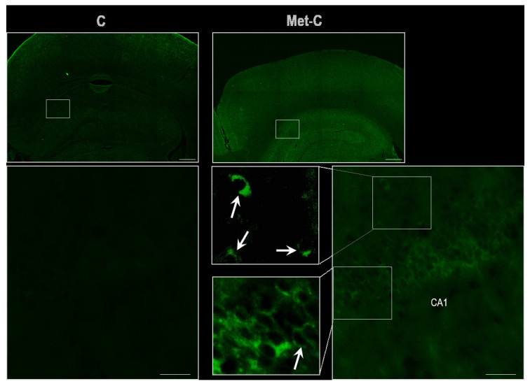
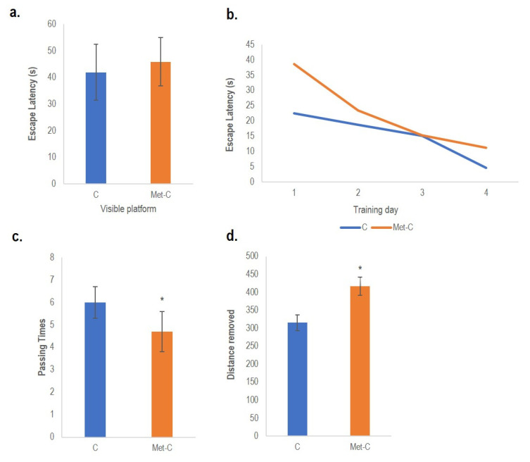
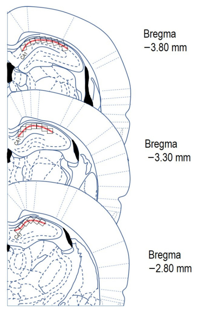
L-methionine, an essential amino acid, plays a critical role in cell physiology. High intake and/or dysregulation in methionine (Met) metabolism results in accumulation of its intermediate(s) or breakdown products in plasma, including homocysteine (Hcy). High level of Hcy in plasma, hyperhomocysteinemia (hHcy), is considered to be an independent risk factor for cerebrovascular diseases, stroke and dementias. To evoke a mild hHcy in adult male Wistar rats we used an enriched Met diet at a dose of 2 g/kg of animal weight/day in duration of 4 weeks. The study contributes to the exploration of the impact of Met enriched diet inducing mild hHcy on nervous tissue by detecting the histo-morphological, metabolomic and behavioural alterations. We found an altered plasma metabolomic profile, modified spatial and learning memory acquisition as well as remarkable histo-morphological changes such as a decrease in neurons' vitality, alterations in the morphology of neurons in the selective vulnerable hippocampal CA 1 area of animals treated with Met enriched diet. Results of these approaches suggest that the mild hHcy alters plasma metabolome and behavioural and histo-morphological patterns in rats, likely due to the potential Met induced changes in "methylation index" of hippocampal brain area, which eventually aggravates the noxious effect of high methionine intake.

Keywords: hyperhomocysteinemia; methionine diet; morris water maze; neurodegeneration; wild-type rats.


Conflict of interest statement

The authors declare no conflict of interest.

Figures

Figure 1
Figure 1
Cresyl violet stained rat brain sections and statistical evaluation of changes in the number of vital neurons in the CA1 region of the hippocampus of the rat brain. (a) Bright-field micrographs of CA1 region of hippocampus representing control (C) and Met-C group–low magnification in the left row, the right row represents high magnification (rectangle) of the corresponding group. Morphologically changed neurons are indicated by a dashed arrow (shrinkage of neurons) and by an asterisk (microvacuolisation and cell swelling), while arrows show vital neurons. CC–corpus callosum, GD–gyrus dentatus. Bar = 200 and 50 μm; n = 5/subgroup. (b) Schematic coronal rat brain section, redrawn according to Paxinos and Watson [31] representing hippocampus (blue rectangle) and CA1 area of the hippocampus (red rectangle). (c) The number of vital neuronal cells in the CA1 area of hippocampus in control and Met-C group. (d) The number of neurons with the marks of degeneration in the CA1 area of hippocampus in control and Met-C group. The significance of group mean differences was evaluated by unpaired t-test. Results are presented as mean ± SD, n = 5/subgroup, *** p < 0.001 versus the control group.
Figure 2
Figure 2
Demonstrative microphotographs of TUNEL staining of rat brain sections in the CA1 region of the hippocampus. Fluorescent micrographs of the hippocampus representing control and Met-C group–low magnification in the first line, the second line represents high magnification (rectangles) of the corresponding group. Arrows indicate perinuclear staining. Bar = 500 and 50 μm; n = 5/subgroup.
Figure 3
Figure 3
Spatial and working memory testing Path length (Distance removed) is shown as an index of spatial learning at the control and Met diet treated animals. Escape latency at the first day of training (a), escape latency during the whole training week on day 2–5. (b), passing times over the platform area (c) and path length spent in the target quadrant after removing the platform (d). Results are presented as mean ± SD for n = 5/subgroup. Behavioural analysis was performed by Student-Newman-Keuls test to compare the means of control (C) and Met-C; * p < 0.05 was considered to be statistically significant.
Figure 4
Figure 4
Schematic representation of three rat brain coronal sections (redrawn and modified according Paxinos and Watson [31]). Red line indicates the area of CA1 where the neurons have been counted. The counting area was divided into the counting grid 0.3 × 0.3 mm (black squares). Each section was counted in the CA1 at least in three of microscopic fields (black squares). Stereotaxic coordinates (from Bregma): −2.80 mm to −3.80 mm.

References

    1. Lehotsky J., Kovalska M., Tomascova A., Kalenska D., Baranovicova E., Kaplan P. Ischemic brain injury in hyperhomocysteinemic conditions and the development of Alzheimer’s disease. In: Pluta R., editor. Brain Ischemia: Alzheimer’s Disease Mechanisms. 1st ed. Nova Science Pub Inc.; New York, NY, USA: 2019. pp. 115–156.
    1. Kovalska M., Hnilicova P., Kalenska D., Tothova B., Adamkov M., Lehotsky J. Effect of Methionine Diet on Metabolic and Histopathological Changes of Rat Hippocampus. Int. J. Mol. Sci. 2019;20:6234. doi: 10.3390/ijms20246234. - DOI - PMC - PubMed
    1. Kovalska M., Tothova B., Kalenska D., Tomascova A., Kovalska L., Adamkov M., Lehotsky J. Association of induced hyperhomocysteinemia with neurodegeneration in rat entorhinal cortex-hippocampal system after global brain ischemia: A progression of Alzheimer’s disease-like pathological features? Act. Nerv. Super. Rediviva. 2019;61:31–38.
    1. Mattson M.P. Gene–Diet Interactions in Brain Aging and Neurodegenerative Disorders. Determinants of Successful Aging: Developing an Integrated Research Agenda for the 21st Century 2. Ann. Intern. Med. 2003;139:441–444. doi: 10.7326/0003-4819-139-5_Part_2-200309021-00012. - DOI - PubMed
    1. Cavallaro R.A., Fuso A., d’Erme M., Miraglia N., Martire S. Role of S-adenosylmethionine in the Modulation of Oxidative Stress-Related Neurodegeneration. Int. J. Clin. Nutr. Diet. 2016;2:109. doi: 10.15344/2456-8171/2016/109. - DOI

LinkOut - more resources