Review
. 2021 May 7;8(2):107-123.
doi: 10.3390/dermatopathology8020017.

Ichthyoses-A Clinical and Pathological Spectrum from Heterogeneous Cornification Disorders to Inflammation

Affiliations
Review

Ichthyoses-A Clinical and Pathological Spectrum from Heterogeneous Cornification Disorders to Inflammation

Dieter Metze et al. Dermatopathology (Basel). .

Abstract

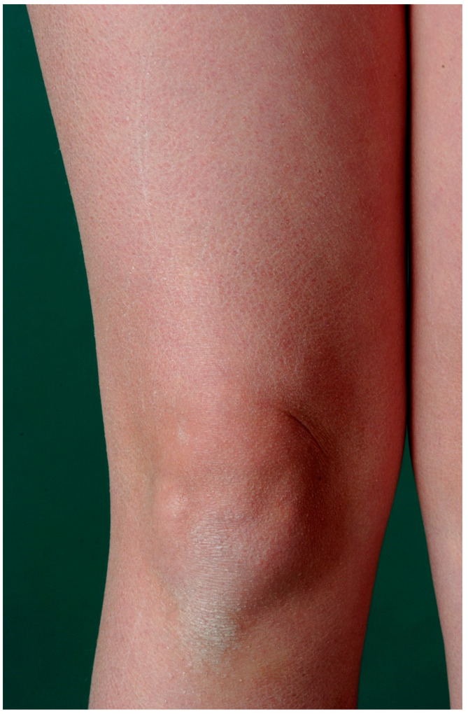
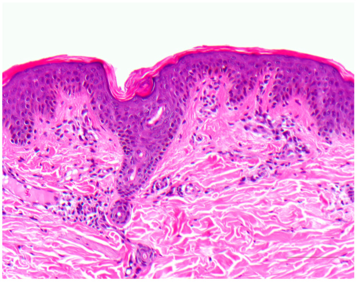
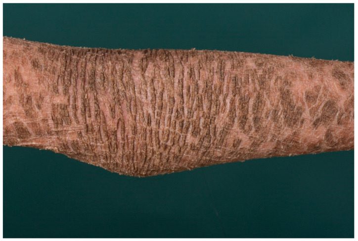
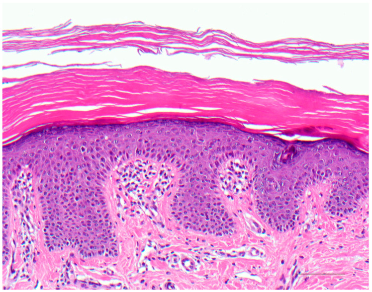
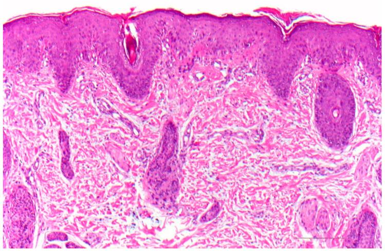
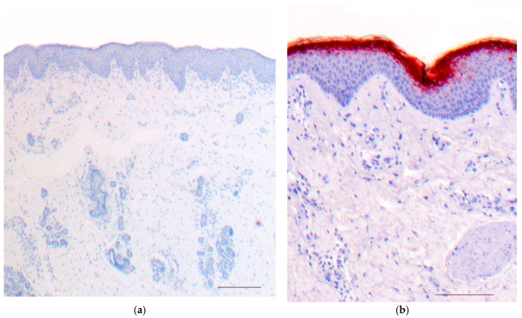
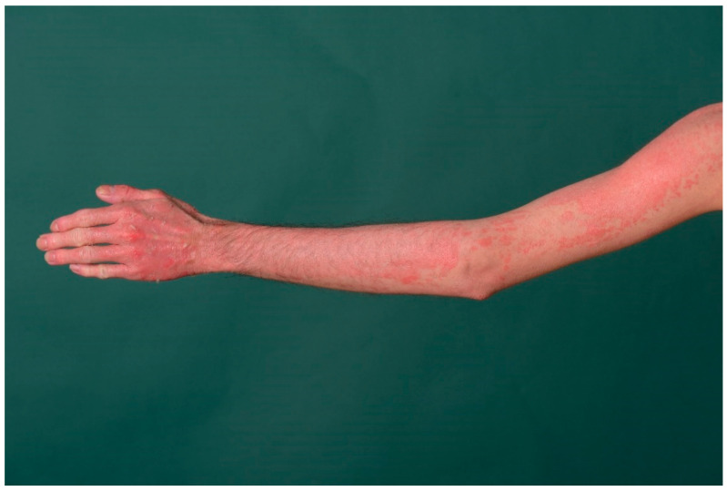
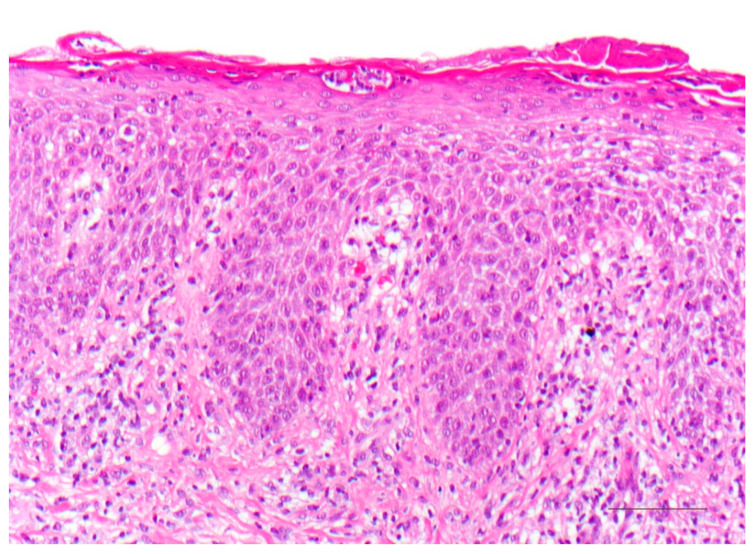
Ichthyoses are inborn keratinization disorders affecting the skin only (non-syndromic) or are associated with diseases of internal organs (syndromic). In newborns, they can be life-threatening. The identification of the gene defects resulted in reclassification and a better understanding of the pathophysiology. Histopathologic patterns include orthohyperkeratosis with a reduced or well-developed stratum granulosum, hyperkeratosis with ortho- and parakeratosis with preserved or prominent stratum granulosum, and epidermolytic ichthyosis. Another pattern features "perinuclear vacuoles and binucleated keratinocytes", which is associated with keratin mutations. Some ichthyoses are histologically defined by psoriasis-like features, and distinct subtypes show follicular hyperkeratosis. In addition to histological and immunohistochemical methods, these patterns allow a better histopathologic diagnosis.

Keywords: dermatopathology; hereditary keratinization disorders; ichthyosis; immunohistochemistry; pattern analysis.


Conflict of interest statement

The authors declare no conflict of interest.

Figures

Figure 1
Figure 1
Ichthyosis vulgaris. Fine grey scaling on the extremities.
Figure 2
Figure 2
Ichthyosis vulgaris. Note the absent stratum granulosum and mild compact orthohyperkeratosis. Marked hyperkeratosis of the opening of the acrosyringium. Inflammatory infiltrates are almost absent. HE stain, bar = 100 µm.
Figure 3
Figure 3
Lamellar ichthyosis. Dark brownish lamellar scaling in a patient with transglutaminase−1 deficiency.
Figure 4
Figure 4
Autosomal recessive lamellar ichthyosis. Acanthotic epidermis with well-developed stratum granulosum and compact orthohyperkeratosis without further signs of inflammation. HE stain, original magnification, bar = 100 µm.
Figure 5
Figure 5
Epidermolytic ichthyosis. Diffuse palmoplantar keratoderma (keratin 1 mutation).
Figure 6
Figure 6
Epidermolytic ichthyosis. Acanthotic epidermis with massive orthohyperkeratosis. Suprabasal keratinocytes vacuolated with distinct hypereosinophilic granules and irregular keratohyalin granules. HE stain, bar = 50 µm.
Figure 7
Figure 7
Erythrokeratoderma. Acanthotic epidermis with orthohyperkeratosis, focal parakeratosis, dyskeratotic keratinocytes, and preserved stratum granulosum. Discrete superficial perivascular lymphocytic infiltrate. HE stain, bar = 100 µm.
Figure 8
Figure 8
KID syndrome (keratitis–ichthyosis–deafness). Dyskeratotic keratinocytes with perinuclear halo (“bird’s eye”). HE stain, bar = 100 µm.
Figure 9
Figure 9
Netherton syndrome. Erythema and scaling of the trunk and face.
Figure 10
Figure 10
Netherton syndrome. Regular (psoriasiform) hyperplasia with focal parakeratosis and thinned stratum granulosum. Dilated vessels in the papillary dermis and inflammatory infiltrates. HE stain, bar = 100 µm.
Figure 11
Figure 11
Netherton syndrome, immunohistochemistry, bar = 200 µm (a,b). Immunohistochemistry shows a lack of staining for LEKTI in the epidermis and hair follicles (a); regular expression of LEKTI in the upper layers of the epidermis of healthy skin, bar = 200 µm (b). Immunoperoxidase staining.
Figure 12
Figure 12
Peeling skin disease. Diffuse erythema with superficial skin detachment is evident from birth and persists throughout life with seasonal variation.
Figure 13
Figure 13
SAM syndrome. Psoriasiform dermatitis with dilated intercellular spaces of the epidermis without blistering (“desmosomal acantholysis”). HE stain, bar = 100 µm.

References

    1. Krug M., Oji V., Traupe H., Berneburg M. Ichthyoses-Part 1: Differential diagnosis of vulgar ichthyoses and therapeutic options. J. Dtsch. Dermatol. Ges. 2009;7:511–519. doi: 10.1111/j.1610-0387.2008.06969.x. - DOI - PubMed
    1. Krug M., Oji V., Traupe H., Berneburg M. Ichthyoses-Part 2: Congenital ichthyoses. J. Dtsch. Dermatol. Ges. 2009;7:577–588. doi: 10.1111/j.1610-0387.2008.06970.x. - DOI - PubMed
    1. Traupe H. A Guide to Clinical Diagnosis, Genetic Counseling, and Therapy. Springer; Berlin, Germany: 1989. The Ichthyoses; pp. 103–138.
    1. Oji V., Traupe H. Ichthyoses: Differential diagnosis and molecular genetics. Eur. J. Dermatol. 2006;16:349–359. - PubMed
    1. Oji V., Tadini G., Akiyama M., Blanchet Bardon C., Bodemer C., Bourrat E., Coudiere P., DiGiovanna J.J., Elias P., Fischer J., et al. Revised nomenclature and classification of inherited ichthyoses: Results of the First Ichthyosis Consensus Conference in Sorèze 2009. J. Am. Acad. Dermatol. 2010;63:607–641. doi: 10.1016/j.jaad.2009.11.020. - DOI - PubMed

LinkOut - more resources