Review
. 2021 May 7;14(5):442.
doi: 10.3390/ph14050442.

The Chemistry Behind ADCs

Affiliations
Review

The Chemistry Behind ADCs

Vesela Kostova et al. Pharmaceuticals (Basel). .

Abstract

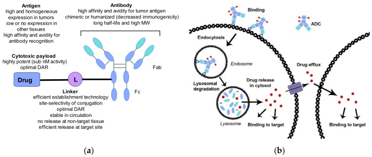
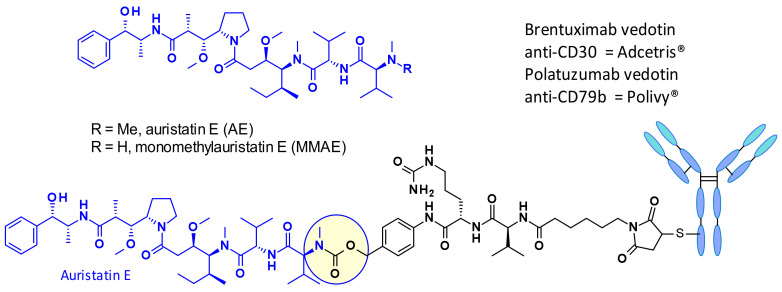
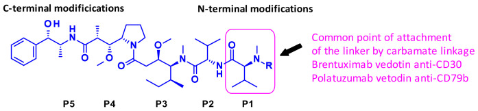
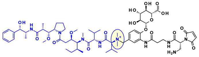
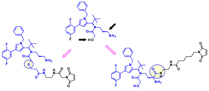
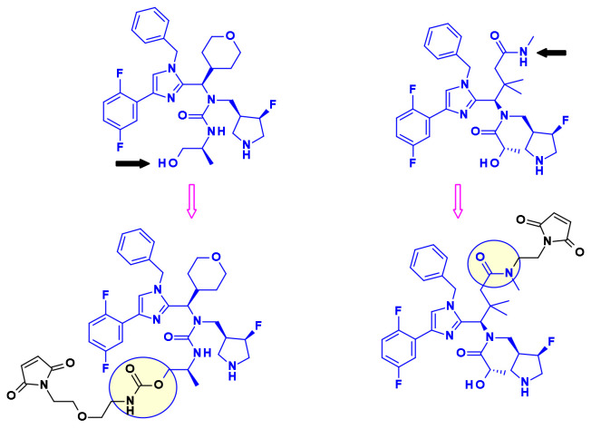
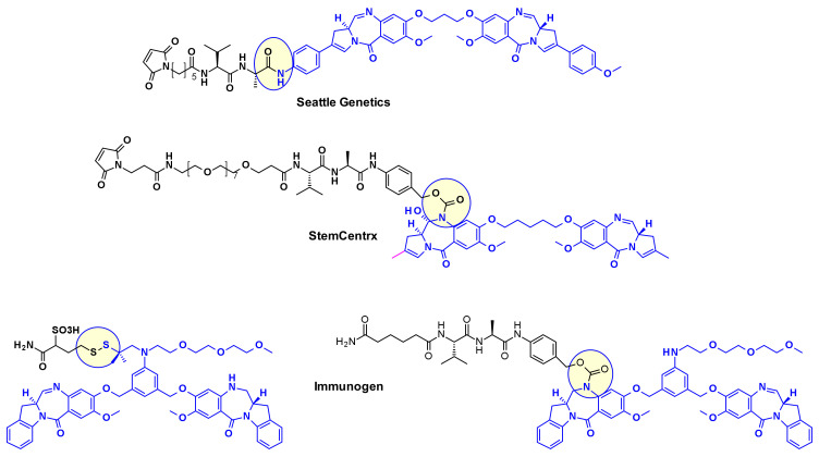
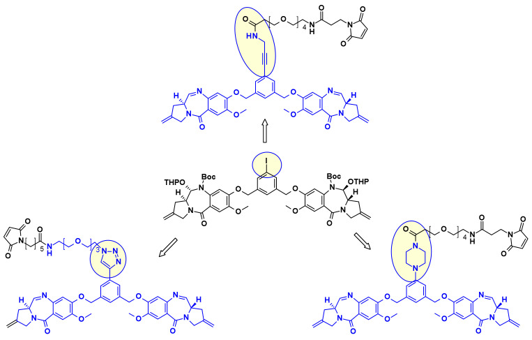
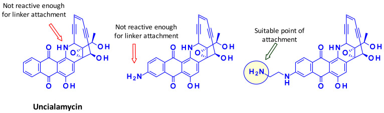
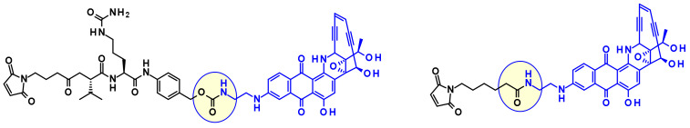
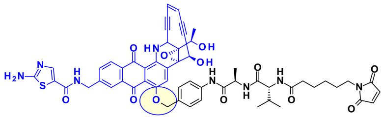
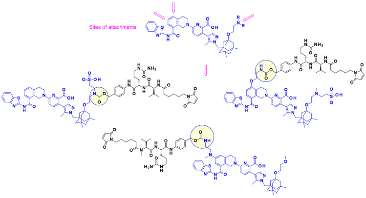
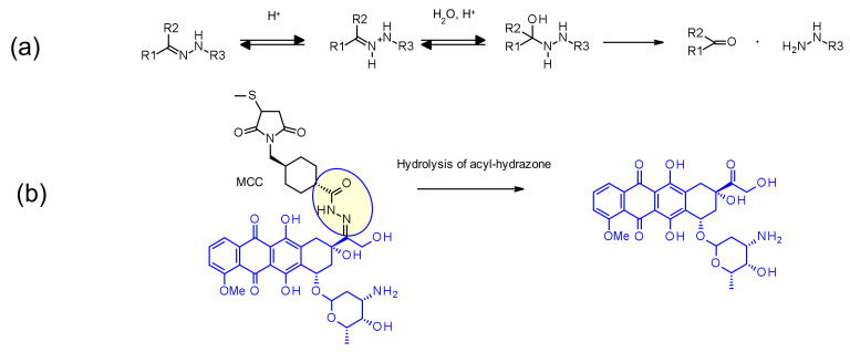
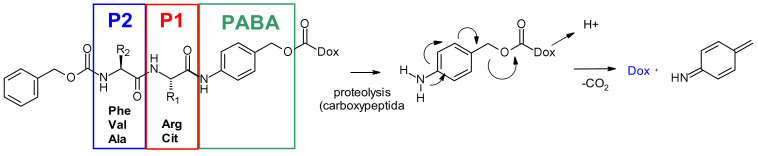
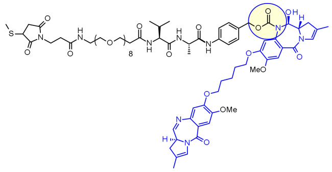
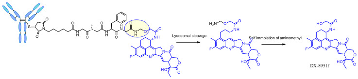
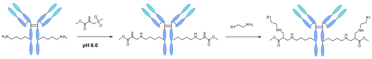
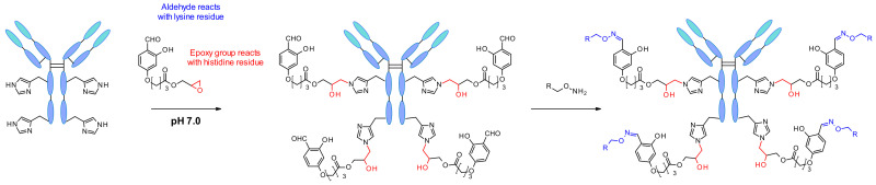
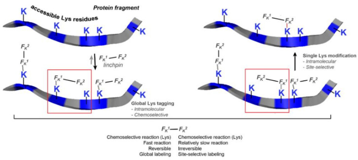
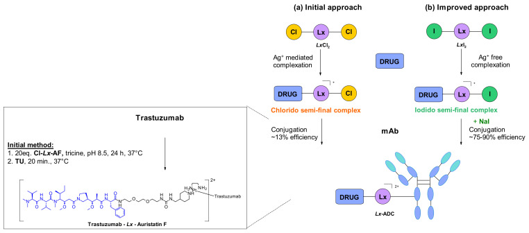
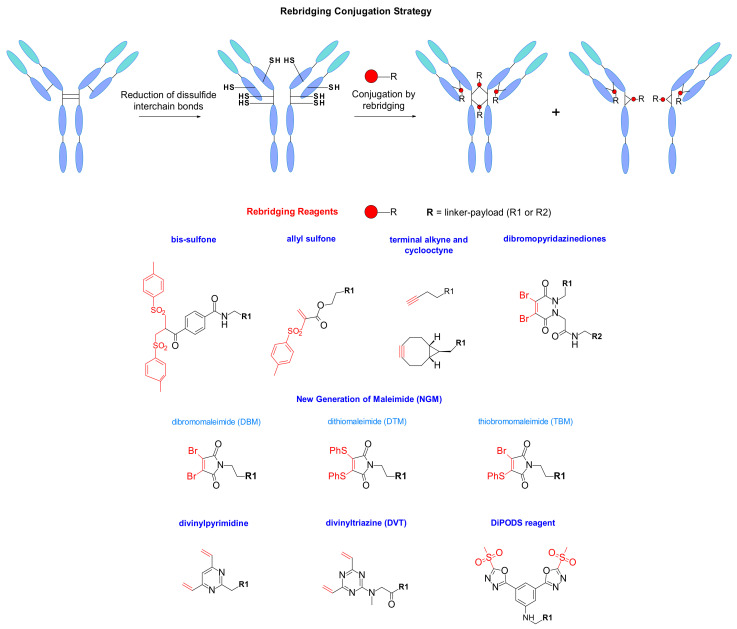
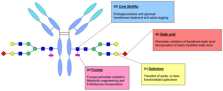
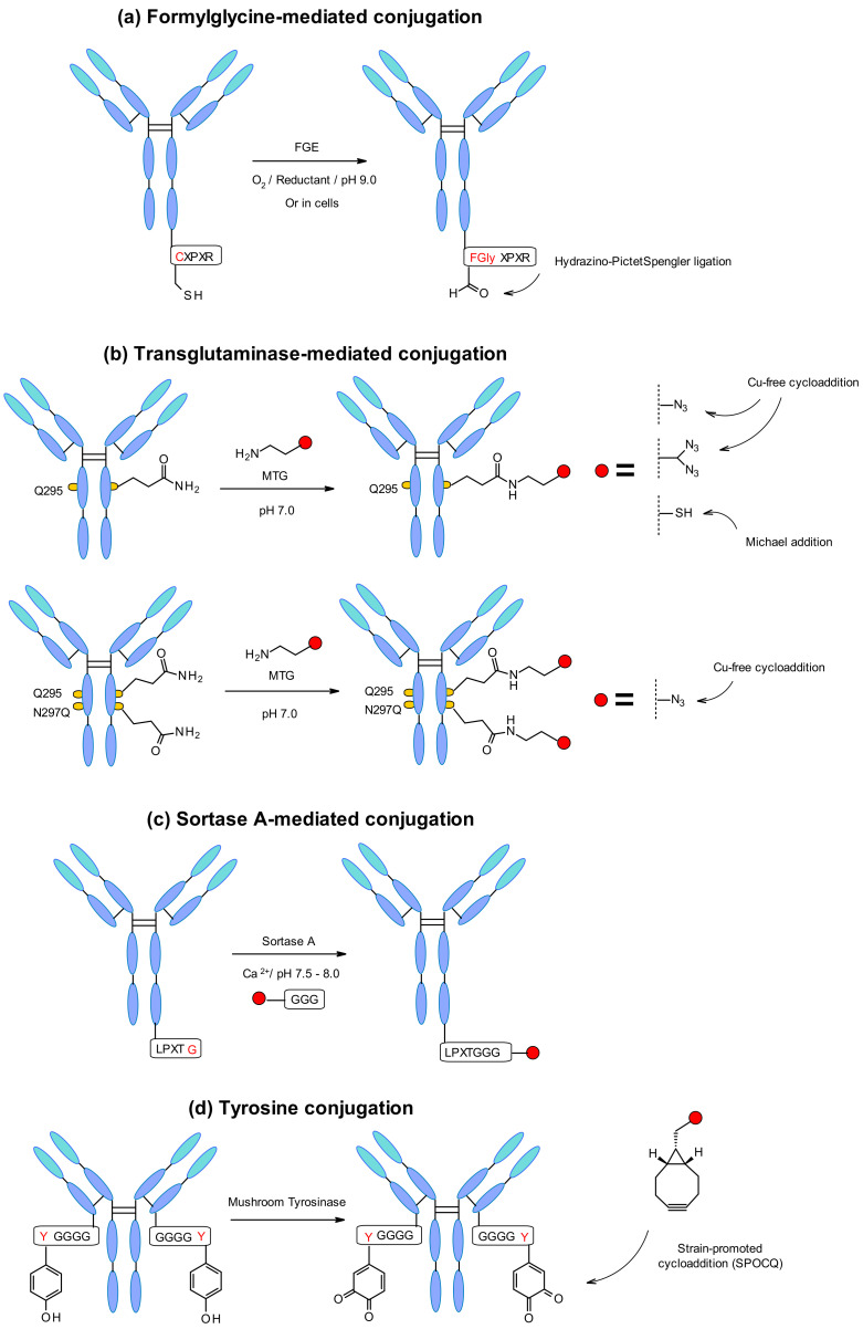
Combining the selective targeting of tumor cells through antigen-directed recognition and potent cell-killing by cytotoxic payloads, antibody-drug conjugates (ADCs) have emerged in recent years as an efficient therapeutic approach for the treatment of various cancers. Besides a number of approved drugs already on the market, there is a formidable follow-up of ADC candidates in clinical development. While selection of the appropriate antibody (A) and drug payload (D) is dictated by the pharmacology of the targeted disease, one has a broader choice of the conjugating linker (C). In the present paper, we review the chemistry of ADCs with a particular emphasis on the medicinal chemistry perspective, focusing on the chemical methods that enable the efficient assembly of the ADC from its three components and the controlled release of the drug payload.

Keywords: antibody-drug conjugates; attachment point; conjugation; linker.


Conflict of interest statement

The authors declare no conflict of interest.

Figures

Figure 1
Figure 1
(a) The general structure of an ADC and the key considerations when combining the different components. (b) Schematic representation of the uptake of the ADC and the release of the payload inside a cancer cell.
Figure 2
Figure 2
Structure of auristatine E (AE), monomethyl auristatin E (MMAE), and commercially approved auristatin-based ADCs: Target Antigen.
Figure 3
Figure 3
Auristatine terminal amino acid sites: N-terminus, P1 and C-terminus, P5.
Figure 4
Figure 4
Seattle Genetics’s linker-payload combination including N-dimethyl auristatine and an ammonium linkage.
Figure 5
Figure 5
Novel drug linkers containing P2 or P4 linkage.
Figure 6
Figure 6
Methylene alkoxy carbamate (MAC) self-immolative unit for alcohol conjugation.
Figure 7
Figure 7
Structure of azastatins.
Figure 8
Figure 8
The structure of maytansine and its conjugable derivatives DM1 and DM4.
Figure 9
Figure 9
Chemical linkage of DM1 and DM4 through a disulfide or sulfide bond.
Figure 10
Figure 10
Maytansine-derived drug-linker precursors.
Figure 11
Figure 11
Structure of Tubulysins and principal points of attachment.
Figure 12
Figure 12
Examples of Tut attached Tubulysin payloads.
Figure 13
Figure 13
Examples of phenyl attached tubulysin linkers payloads.
Figure 14
Figure 14
Examples of Tubulysin payloads attached to linkers through the Mep group.
Figure 15
Figure 15
Two approaches for introducing handle in Cryptomycin.
Figure 16
Figure 16
Examples of cryptophycin-derived linker-payload precursors.
Figure 17
Figure 17
Identification of linker attachment points of the pyrrole based KSP inhibitor and examples of linker-payload conjugates prepared by Bayer.
Figure 18
Figure 18
Vectorization of imidazole-based KSP inhibitors.
Figure 19
Figure 19
Attachment of linkers to PBD and IBD payloads.
Figure 20
Figure 20
PBD dimer/linkers attachment using a common iodobenzene intermediate.
Figure 21
Figure 21
Activation of duocamycin derivatives and mechanism of action of alkylation of N3 adenine in the minor groove of DNA.
Figure 22
Figure 22
Structure of the ADCs developed by Synthon (SYD985) and Medarex (BMS9336561).
Figure 23
Figure 23
Structure of camptothecin, irinotecan and its metabolite SN-38.
Figure 24
Figure 24
The vectorization points of SN38 and representative linker-payload conjugates.
Figure 25
Figure 25
Structures of exatecan and its derivatives, DXd(1) and DXd(2), as well as the derived conjugates.
Figure 26
Figure 26
Elimination of the ring F by Immunogen and design of new related camptothecin L/P.
Figure 27
Figure 27
Structure of calicheamicins and the design of the two marketed ADCs Mylotarg® and Besponsa®.
Figure 28
Figure 28
Mechanism of di radical formation from activated calicheamicin.
Figure 29
Figure 29
Research of a suitable handle on Uncialamycin.
Figure 30
Figure 30
Cleavable and non-cleavable linker-payload conjugates.
Figure 31
Figure 31
Linking of an uncialamycin analog through its phenol group.
Figure 32
Figure 32
Attachment sites on a typical Bcl-xL inhibitor and some reported payload-linker conjugates.
Figure 33
Figure 33
Structure of thailanstatin A.
Figure 34
Figure 34
Unexpected Diels Alder reaction between the diene present in the thailanstatine structure and bioconjugation motif, maleimide.
Figure 35
Figure 35
Thailanstatine-based payload-linker conjugate bearing amine spacer on the carboxylic acid and an iodo acetamide end group to avoid intramolecular cyclisation.
Figure 36
Figure 36
Pfizer’s “linker less” thailanstatin ADC.
Figure 37
Figure 37
(a) Structures of the bicyclic octapeptide toxins α-amanitin and β-amanitin and their amino acid constituent numbering (b) Conjugation sites available in amatoxin for coupling to antibodies through linkers
Figure 38
Figure 38
Structure of Heidelberg Pharma’s ADC precursor.
Figure 39
Figure 39
Structure of an OHPAS-linked α-Amanitin payload.
Figure 40
Figure 40
NAMPT inhibitors as payloads for ADCs.
Figure 41
Figure 41
Structure of carmaphycin A and B.
Figure 42
Figure 42
Evolution of carmaphycin B in order to install a suitable amine handle and examples of non-cleavable and cleavable ADCs precursors.
Figure 43
Figure 43
Structure of trastuzumab emtansine (Kadcyla®).
Figure 44
Figure 44
(a) Schematic structure of disulfide-containing linker and its reaction with thiols, like GSH to release payload. (b) Schematic representation of maytansinoid ADC linker highlighting the role of alpha-methyl groups (R1 to R4). (c) Linker structure of SAR-3419 containing the DM4 payload and the SPDB linker.
Figure 45
Figure 45
(a) Hydrolysis of hydrazone in acidic conditions (b) Cleavage of acyl hydrazone linker present in IMMU-110, releasing doxorubicin.
Figure 46
Figure 46
Release mechanism from the dipeptide-PABC-doxorubicin conjugate. Doxorubicin, linked by its primary amine, is not present for clarity.
Figure 47
Figure 47
Chemical structure of Loncastuximab tesirine.
Figure 48
Figure 48
Chemical structure of trastuzumab deruxtecan.
Figure 49
Figure 49
Schematic structure and postulated release mechanism of phosphate (n = 1) and pyrophosphate (n = 2) linkers. Conjugation type to antibody and payload (budesonide) not represented for clarity. Budesonide is linked by its primary alcohol.
Figure 50
Figure 50
Chemical structure and release mechanism of pyrophosphate diester linkers.
Figure 51
Figure 51
(a) Chemical structure of β-glucuronic linker and corresponding release mechanism. Drugs contains primary (doxorubicin) or secondary amine (MMAE, MMAF) (b) Chemical structure and release mechanism of DMED-containing β-Glucuronidase cleavable linker. Drug structures are omitted for clarity.
Figure 52
Figure 52
Chemical structure of β-galactosidase linker and mechanism of release.
Figure 53
Figure 53
Chemical structure of sulfatase-cleaved linker and the corresponding release mechanism. Both drug structure and antibody omitted for clarity (rebridging conjugation).
Figure 54
Figure 54
Sulfonyl acrylate reagent reacting with lysine residues of native mAb.
Figure 55
Figure 55
Linchpin directed conjugation on histidine residues performed by multitasking group reagent.
Figure 56
Figure 56
Hypothetic mechanism of linchpin technology for site-specific conjugation on lysine residues based on Fk1-spacer-Fk2 reagent.
Figure 57
Figure 57
Site-specific conjugation approach on histidine residues by cationic metal-organic PtII-based linker: (a) initial approach generating chlorido semi-final complex. (b) Improved approach through iodido semi-final complex.
Figure 58
Figure 58
Rebridging conjugation strategy for modification of native antibodies using rebridging reagents.
Figure 59
Figure 59
(a) Fucose modification by oxidation with periodate or by metabolic engineering of thiolated analogue. (b) Enzymatic addition of terminal sialic acids followed by periodate oxidation or incorporation of azido modified sialic acid. (c) Incorporation of azido- or keto- functionalized galactose. (d) Endoglycosidase homogenizing the glycan structure and incorporation of an azide anchor enabling copper-free cycloaddition.
Figure 60
Figure 60
(a) Cysteine residue in pentapeptide Cys-X-Pro-X-Arg oxidized to formylglycine by FGE generating bioothogonal handle. (b) Microbial transglutaminase (MTGase) strategy on glutamine at position 295 and improved approach by incorporation of additional mutation N297Q. (c) Sortase-mediated conjugation exploiting glycine-functionalised payloads. (d) Tyrosine oxidation by mushroom tyrosinase generating biorthogonal handle for strain-promoted cycloaddition. For clarity, the reaction (a) and (c) is only depicted in one heavy chain.

References

    1. Drago J.Z., Modi S., Chandarlapaty S. Unlocking the potential of antibody–drug conjugates for cancer therapy. Nat. Rev. Clin. Oncol. 2021 doi: 10.1038/s41571-021-00470-8. - DOI - PMC - PubMed
    1. Khongorzul P., Ling C.J., Khan F.U., Ihsan A.U., Zhang J. Antibody–Drug Conjugates: A Comprehensive Review. Mol. Cancer Res. 2020;18:3–19. doi: 10.1158/1541-7786.MCR-19-0582. - DOI - PubMed
    1. Joubert N., Beck A., Dumontet C., Denevault-Sabourin C. Antibody–Drug Conjugates: The Last Decade. Pharmaceuticals. 2020;13:245. doi: 10.3390/ph13090245. - DOI - PMC - PubMed
    1. Ritchie M., Tchistiakova L., Scott N. Implications of receptor-mediated endocytosis and intracellular trafficking dynamics in the development of antibody drug conjugates. mAbs. 2013;5:13–21. doi: 10.4161/mabs.22854. - DOI - PMC - PubMed
    1. O’Sullivan M.J., Lindsay A.J. The Endosomal Recycling Pathway—At the Crossroads of the Cell. Int. J. Mol. Sci. 2020;21:6074. doi: 10.3390/ijms21176074. - DOI - PMC - PubMed

LinkOut - more resources