Review
. 2021 May 7;10(9):2008.
doi: 10.3390/jcm10092008.

Hyperferritinemia-A Clinical Overview

Affiliations
Review

Hyperferritinemia-A Clinical Overview

Miriam Sandnes et al. J Clin Med. .

Abstract

Ferritin is one of the most frequently requested laboratory tests in primary and secondary care, and levels often deviate from reference ranges. Serving as an indirect marker for total body iron stores, low ferritin is highly specific for iron deficiency. Hyperferritinemia is, however, a non-specific finding, which is frequently overlooked in general practice. In routine medical practice, only 10% of cases are related to an iron overload, whilst the rest is seen as a result of acute phase reactions and reactive increases in ferritin due to underlying conditions. Differentiation of the presence or absence of an associated iron overload upon hyperferritinemia is essential, although often proves to be complex. In this review, we have performed a review of a selection of the literature based on the authors' own experiences and assessments in accordance with international recommendations and guidelines. We address the biology, etiology, and epidemiology of hyperferritinemia. Finally, an algorithm for the diagnostic workup and management of hyperferritinemia is proposed, and general principles regarding the treatment of iron overload are discussed.

Keywords: ferritin; hemochromatosis; inflammation; iron.


Conflict of interest statement

The authors declare no conflict of interest.

Figures

Figure 1
Figure 1
Schematic overview of regulated ferritin translation by the iron responsive element (IRE)/iron regulatory protein 1 (IRP1) system. Upon low intracellular iron levels (A), IRP1 binds to IRE, inhibiting the recruitment of the small ribosomal unit to mRNA, which results in a small ferritin pool. Upon high intracellular iron levels (B), iron binds to IRP1, causing a transformational change that dissociates IRP1 from IRE. Both ribosomal subunits are now recruited to the mRNA and the translation of ferritin is activated. This coordinated IRP1/IRE binding with respect to iron levels stabilizes cellular iron through the synthesis of ferritin.
Figure 2
Figure 2
Body iron homeostasis and the hepcidin–ferroportin axis. Dietary non-heme is absorbed at the apical site of enterocytes of the jejunum through divalent metal transporter 1 (DMT1). One portion remains stored as ferritin inside the enterocyte, while the rest is transferred through the basolateral site via ferroportin. Iron subsequently binds transferrin and is further distributed throughout the body. Most iron is distributed to bone marrow for hemoglobin production. Senescent erythrocytes are phagocytosed by macrophages of the reticuloendothelial system (RES), and iron is catabolized from hemoglobin before subsequently re-entering circulation. Hepcidin produced in the liver regulates the systemic iron balance through binding to ferroporrtin. This binding facilitates the lysosomal degradation of the iron exporter, ultimately resulting in decreased serum iron concentrations.
Figure 3
Figure 3
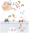
Schematic overview of hereditary hemochromatosis (HH) associated with impaired hepcidin expression. HFE disease-associated variants (type 1 HH) cause impaired assembly of the iron sensing complex (A), as transferrin binding to transferrin receptor 1 (TfR1) normally competes with and releases HFE to interact with transferrin receptor 2 (TfR2). Disease-associated variants of the HJV (type 2A HH) and TFR2 (type 3 HH) genes, encoding subunits of the iron sensing complex, cause impaired function of this complex which normally regulates hepcidin transcription through a signaling cascade (B). Disease-associated variants of the HAMP (hepcidin) gene (type 2B HH) cause reduced hepcidin levels, despite a functioning iron sensing complex (C). An impaired hepcidin–ferroportin axis (D) results in uncontrolled intestinal iron absorption and the release of iron stores from macrophages and parenchymal cells, and, accordingly, high serum iron levels. When transferrin saturation is highly elevated, the transferrin iron-binding capacity is exceeded and non-transferrin bound iron (NTBI) enters circulation. NTBI is a more toxic form than transferrin-bound iron, capable of producing reactive oxygen species, which are involved in the cellular damage seen upon iron-loading conditions, for example, liver fibrosis and cirrhosis. NTBI is eliminated from circulation through transporters mainly expressed on hepatocytes (E). Here, it is subsequently stored in iron-storing complexes such as ferritin and hemosiderin to limit cellular damage.
Figure 4
Figure 4
Variants of the FPN1 (SLC40A1) gene encoding the iron exporter ferroportin involved in type 4 hereditary hemochromatosis (HH). Loss-of-function variants (A) impair the iron-export capability or expression of ferroportin, leading to iron accumulation mainly in cells such as tissue macrophages and discrete accumulation in parenchymal cells, decreased iron delivery to circulating transferrin causing an inappropriately low transferrin saturation, and decreased iron delivery for hemoglobin production. This is referred to as ferroportin disease, or type 4A HH. Gain-of-function variants (B), however, make ferroportin resistant to hepcidin-induced degradation, which normally inhibits the export of iron. This results in a similar impairment of the hepcidin–ferroportin axis, as seen in type 1-3 HH, with high serum iron concentrations and transferrin saturation, and iron mainly accumulating in parenchymal cells and hepatocytes. This is often referred to as type 4B HH.
Figure 5
Figure 5
Diagnostic workup and management in hyperferritinemia of unknown cause. CRP: C-reactive protein; ESR: erythrocyte sedimentation rate; MRI, magnetic resonance imaging.

Similar articles

Cited by

References

    1. Cullis J.O., Fitzsimons E.J., Griffiths W.J., Tsochatzis E., Thomas D.W., Haematology T.B.S.F. Investigation and management of a raised serum ferritin. Br. J. Haematol. 2018;181:331–340. doi: 10.1111/bjh.15166. - DOI - PubMed
    1. Jacobs A., Miller F., Worwood M., Beamish M.R., Wardrop C.A. Ferritin in the serum of normal subjects and patients with iron deficiency and iron overload. Br. Med. J. 1972;4:206–208. doi: 10.1136/bmj.4.5834.206. - DOI - PMC - PubMed
    1. Knovich M.A., Storey J.A., Coffman L.G., Torti S.V., Torti F.M. Ferritin for the clinician. Blood Rev. 2009;23:95–104. - PMC - PubMed
    1. Guyatt G.H., Oxman A.D., Ali M., Willan A., McIlroy W., Patterson C. Laboratory diagnosis of iron-deficiency anemia: An overview. J. Gen. Intern. Med. 1992;7:145–153. doi: 10.1007/BF02598003. - DOI - PubMed
    1. Beaton M.D., Adams P.C. Treatment of hyperferritinemia. Ann. Hepatol. 2012;11:294–300. - PubMed

LinkOut - more resources