Review
. 2021 May 22;22(11):5461.
doi: 10.3390/ijms22115461.

Transmembrane TNF and Its Receptors TNFR1 and TNFR2 in Mycobacterial Infections

Affiliations
Review

Transmembrane TNF and Its Receptors TNFR1 and TNFR2 in Mycobacterial Infections

Andy Ruiz et al. Int J Mol Sci. .

Abstract

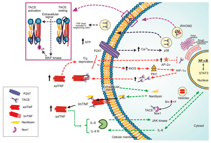
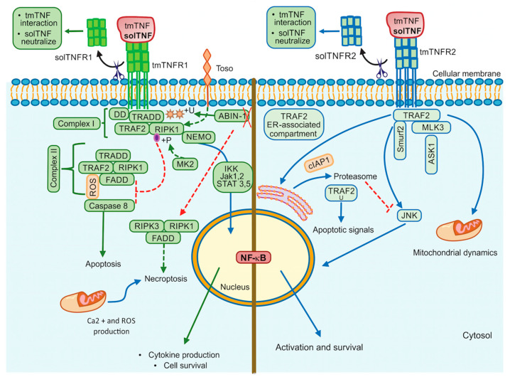
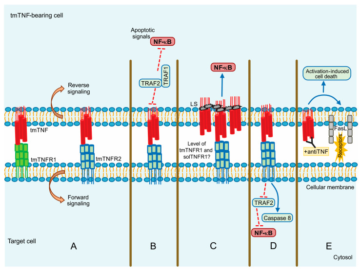
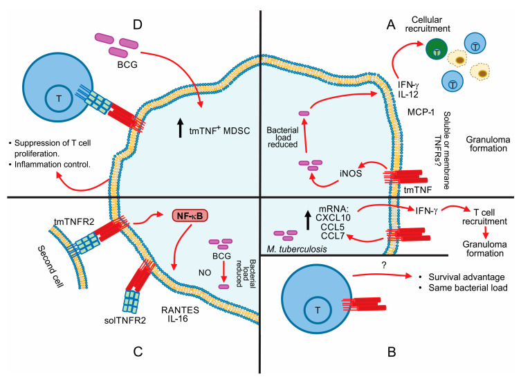
Tumor necrosis factor (TNF) is one of the main cytokines regulating a pro-inflammatory environment. It has been related to several cell functions, for instance, phagocytosis, apoptosis, proliferation, mitochondrial dynamic. Moreover, during mycobacterial infections, TNF plays an essential role to maintain granuloma formation. Several effector mechanisms have been implicated according to the interactions of the two active forms, soluble TNF (solTNF) and transmembrane TNF (tmTNF), with their receptors TNFR1 and TNFR2. We review the impact of these interactions in the context of mycobacterial infections. TNF is tightly regulated by binding to receptors, however, during mycobacterial infections, upstream activation signalling pathways may be influenced by key regulatory factors either at the membrane or cytosol level. Detailing the structure and activation pathways used by TNF and its receptors, such as its interaction with solTNF/TNFRs versus tmTNF/TNFRs, may bring a better understanding of the molecular mechanisms involved in activation pathways which can be helpful for the development of new therapies aimed at being more efficient against mycobacterial infections.

Keywords: TNF receptors; mycobacterial infections; tumor necrosis factor; tumor necrosis factor-α converting enzyme (TACE).


Conflict of interest statement

The authors declare no conflict of interest.

Figures

Figure 1
Figure 1
Regulation pathways to the TNF/TACE axis, ways to lead the TACE activation. Dotted green line: Nardilysin-dependent pathways. Dotted red line: iNOS/AP-2a-dependent pathways. +P Phosphorylate form. Dotted blue black line: P2X7 dependent pathway, Dotted purple line: iRHOM2 as the TACE maturation promoter. Black arrow up: increased level.
Figure 2
Figure 2
TNFR1 and TNFR2 activation pathways. TNFR1 signalling is involved in cell death events and inflammatory processes by classical NF-kB, whereas TNRF2 triggers signalling that may activate classical or non-classical NF-kB pathway. Additionally, TNFR2 impacts mitochondrial dynamics. ABIN-1: Ubiquitin binding protein, DD: Death domain, FADD: Fas-associated death domain, IKK: IkappaB kinase, Jak: Janus kinase protein, NEMO: The NF-κB essential modulator (or IKKγ), RIPK1: Receptor interacting protein kinase 1, RIPK3: Receptor interacting protein kinase 3, ROS: Reactive oxygen species, STAT: Signal transducer and activator of transcription proteins, Toso: Human Fcμ receptor (hFCMR) or FAIM3, TRADD: TNFR1-associated death domain, TRAF2: TNFR-associated factor 2, +P: Phosphorylation, +U: Ubiquitination. Continuous lines green and blue: activation signalling. Dotted lines: inhibition signalling. A red cross: absence.
Figure 3
Figure 3
tmTNF signalling pathway. (A) Forward and reverse signalling; (B) Apoptotic signal by TRAF1 and TRAF2; (C) Direct NF-kB activation by LS (leader sequence) in tmTNF; (D) Actin involvement in tm-TNF transduction; (E) Activation Induced Cell Death (AICD) by upregulation of FASL (CD95L). Continuous blue lines: activation signalling. Dotted red lines: inhibition signalling.
Figure 4
Figure 4
tmTNF in mycobacterial infection. (A) tmTNF and iNOS in granulomas promote the secretion of Th1 chemokines and cytokines; (B) Up: M. tuberculosis acute-phase infection in mice. Below: tmTNF expression in T cells during M. tuberculosis infection. (C) tmTNF regulation by tmTNFR2 or solTNFR2; (D) tmTNF expression on MDSC (Myeloid-Derived Suppressor Cells) leads to specific interaction with tmTNFR2. Black arrow up: increased level.

References

    1. Müller U., Jongeneel C.V., Nedospasov S.A., Lindahl K.F., Steinmetz M. Tumour Necrosis Factor and Lymphotoxin Genes Map Close to H-2D in the Mouse Major Histocompatibility Complex. Nature. 1987;325:265–267. doi: 10.1038/325265a0. - DOI - PubMed
    1. Spies T., Morton C.C., Nedospasov S.A., Fiers W., Pious D., Strominger J.L. Genes for the Tumor Necrosis Factors Alpha and Beta Are Linked to the Human Major Histocompatibility Complex. Proc. Natl. Acad. Sci. USA. 1986;83:8699–8702. doi: 10.1073/pnas.83.22.8699. - DOI - PMC - PubMed
    1. Brehm M.A., Daniels K.A., Welsh R.M. Rapid Production of TNF-Alpha Following TCR Engagement of Naive CD8 T Cells. J. Immunol. 2005;175:5043–5049. doi: 10.4049/jimmunol.175.8.5043. - DOI - PubMed
    1. Wingfield P., Pain R.H., Craig S. Tumour Necrosis Factor Is a Compact Trimer. FEBS Lett. 1987;211:179–184. doi: 10.1016/0014-5793(87)81432-1. - DOI - PubMed
    1. Chensue S.W., Remick D.G., Shmyr-Forsch C., Beals T.F., Kunkel S.L. Immunohistochemical Demonstration of Cytoplasmic and Membrane-Associated Tumor Necrosis Factor in Murine Macrophages. Am. J. Pathol. 1988;133:564–572. - PMC - PubMed

MeSH terms

Substances