Review
. 2021 May 22;11(6):779.
doi: 10.3390/biom11060779.

Mechanisms That Activate 26S Proteasomes and Enhance Protein Degradation

Affiliations
Review

Mechanisms That Activate 26S Proteasomes and Enhance Protein Degradation

Alfred L Goldberg et al. Biomolecules. .

Abstract

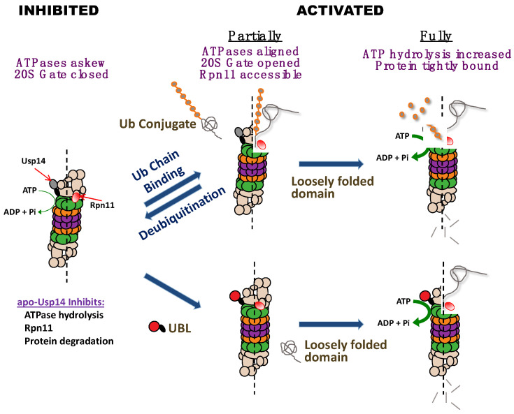
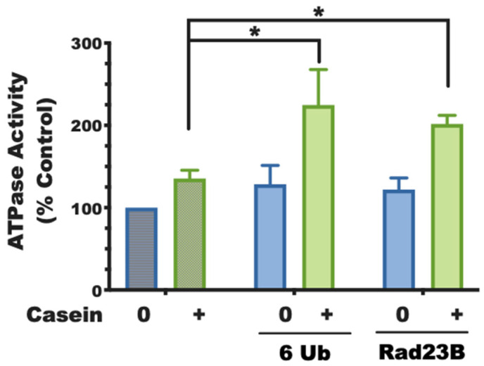
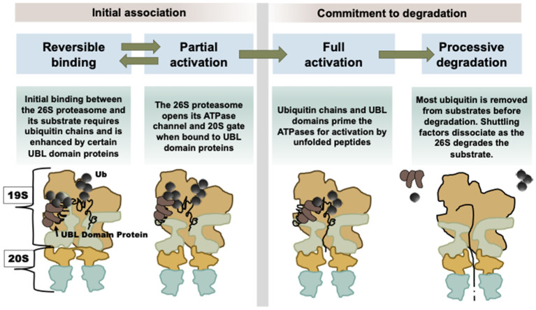
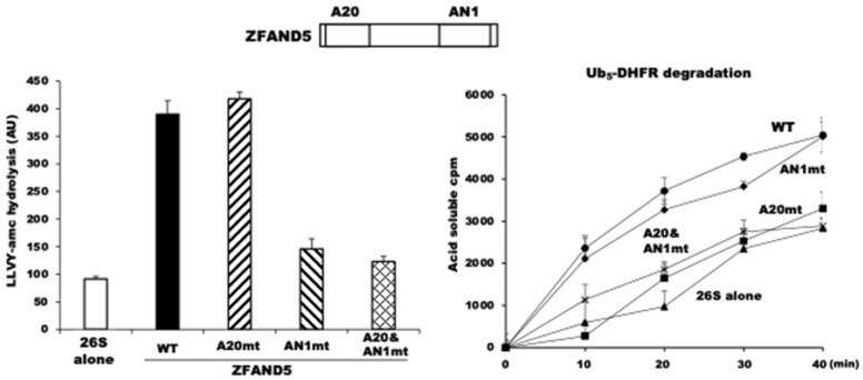
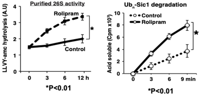
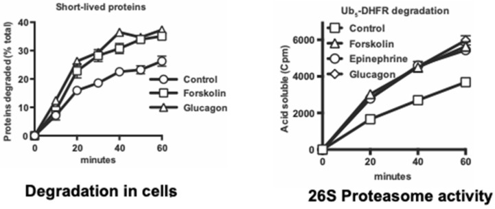
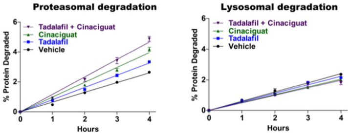
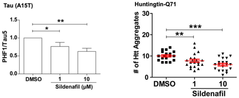
Although ubiquitination is widely assumed to be the only regulated step in the ubiquitin-proteasome pathway, recent studies have demonstrated several important mechanisms that regulate the activities of the 26S proteasome. Most proteasomes in cells are inactive but, upon binding a ubiquitinated substrate, become activated by a two-step mechanism requiring an association of the ubiquitin chain with Usp14 and then a loosely folded protein domain with the ATPases. The initial activation step is signaled by Usp14's UBL domain, and many UBL-domain-containing proteins (e.g., Rad23, Parkin) also activate the proteasome. ZFAND5 is a distinct type of activator that binds ubiquitin conjugates and the proteasome and stimulates proteolysis during muscle atrophy. The proteasome's activities are also regulated through subunit phosphorylation. Agents that raise cAMP and activate PKA stimulate within minutes Rpn6 phosphorylation and enhance the selective degradation of short-lived proteins. Likewise, hormones, fasting, and exercise, which raise cAMP, activate proteasomes and proteolysis in target tissues. Agents that raise cGMP and activate PKG also stimulate 26S activities but modify different subunit(s) and stimulate also the degradation of long-lived cell proteins. Both kinases enhance the selective degradation of aggregation-prone proteins that cause neurodegenerative diseases. These new mechanisms regulating proteolysis thus have clear physiological importance and therapeutic potential.

Keywords: PKA; PKG; Rad23; UBL-domain-containing proteins; Usp14; ZFAND5; ubiquitin–proteasome system.


Conflict of interest statement

The authors declare no conflict of interest.

Figures

Figure 1
Figure 1
26S proteasomes inhibited by Usp14 become activated either upon ubiquitin conjugate binding to Usp14 or upon binding another cell protein containing a UBL domain. In the absence of a ubiquitin conjugate, Usp14 inhibits several proteasome activities: peptide hydrolysis (due to the misalignment of ATPases and the closed 20S gate), ATP hydrolysis, Rpn11-mediated de-ubiquitination, and consequently protein degradation. However, upon binding to a ubiquitin conjugate, Usp14, through its UBL domain, activates proteasomes: the ATPases align, the 20S gate opens, and Rpn11 becomes accessible to substrates. If a loosely folded domain is also present in the substrate, proteasomes become fully active, ATP hydrolysis increases, and the protein substrate is bound more tightly, leading to processive degradation. Alternatively, the binding to the proteasome of a protein containing a UBL domain (e.g., a UBL-UBA shuttling factor) stimulates peptide hydrolysis. Full activation occurs if, in addition, there is present a protein with a loosely folded domain.
Figure 2
Figure 2
Activation of the 26S ATPases requires two signals. The basal rate of ATP hydrolysis by rabbit muscle proteasomes can be stimulated by a ubiquitinated substrate provided it contains both a ubiquitin chain (e.g., here, a linear hexa-ubiquitin, 6 Ub) and a protein with a loosely folded domain (e.g., casein). Normally, these two features are covalently linked in a ubiquitinated substrate but, as shown here, they can activate if on separate molecules. Additionally, isolated UBL domains or a full-length UBL protein (e.g., Rad23B) can replace ubiquitin chains in activating the 26S ATPases (adapted from [8]). (Mean of three experiments ± SEM; * denotes p < 0.05).
Figure 3
Figure 3
Our present understanding of how UBL-UBA shuttling factors stimulate proteasomal degradation of client proteins. This mechanism summarizes multiple steps leading to processive degradation. Shuttling factors (Rad23A, Rad23B, Ddi2, Ubqln1, Ubqln2, and Ubqln4) facilitate the delivery of ubiquitinated proteins to proteasomes and, with their UBL domains, trigger this multistep activation mechanism, which involves stimulating the ATPases (the light green objects in the 19S particle) and opening the gates of the 20S α-subunits (the orange objects in the 20S particle) (for further discussion, see [31]).
Figure 4
Figure 4
ZFAND5 directly activates purified 26S proteasomes and increases their ability to hydrolyze peptides and ubiquitin conjugates. (Left Panel) ZFAND5 stimulates the peptidase activity of 26S proteasomes. This activation is dependent on ZFAND5’s C-terminal AN1 domain. (Right Panel) ZFAND5 enhances also degradation of ubiquitinated 32P-dehydrofolate reductase. Its A20 domain is essential for the increased degradation of ubiquitinated proteins.
Figure 5
Figure 5
26S proteasomes from mouse myotubes treated with the PDE4 inhibitor, rolipram, are more active in hydrolyzing peptides and a ubiquitinated protein. C2Cl2 myotubes were incubated with rolipram, and, at different times, samples were taken and 26S proteasomes purified by the UBL method (Left Panel). Chymotrypsin-like peptidase activity was increased in the treated cells. (Right Panel) After 6 h treatment of cells with rolipram, the purified proteasomes show a greater capacity to degrade ubiquitinated 32P-Sic1 (adapted from [15]). (Mean of three experiments ± SEM; * denotes p < 0.01).
Figure 6
Figure 6
In mouse hepatocytes, glucagon and epinephrine, like forskolin, rapidly stimulate breakdown of short-lived proteins and the proteasomes’ ability to degrade ubiquitinated substrates. (Left Panel) Short-lived proteins in primary mouse hepatocytes were initially labeled for 20 min with 3H-phenylanine, as described previously [62,64]. After labeling and re-suspension in chase medium, degradation was measured in the presence of glucagon or forskolin. (Right Panel) 26S proteasomes were purified by the UBL method from (nonlabeled) mouse hepatocytes treated for 1 h with either forskolin, epinephrine, glucagon, or the vehicle. These treatments increased the ability of proteasomes to degrade the model UPS substrate, ubiquitinated 32P-labeled dihydrofolate reductase (adapted from [16]).
Figure 7
Figure 7
Agents that raise cGMP increase the degradation of proteins by the UPS, but not by autophagy. Proteins in SY5Y cells were prelabeled with 3H-phenylanine for 20 h to label long-lived cell proteins as described previously [62,64]. Proteasomal degradation reflects the net decrease in total protein breakdown in the presence of the proteasome inhibitor bortezomib. Lysosomal degradation represents the net decrease in the presence of the inhibitor of lysosome acidification concanamycin A (adapted from [16]).
Figure 8
Figure 8
Raising cGMP levels promotes the clearance of mutant proteins in zebrafish models of a tauopathy and Huntington’s disease. (Left Panel) Zebrafish larvae expressing the tauopathy-associated A15T mutation in neurons were treated with sildenafil for 5 days. Their content of the hyperphosphorylated tau, expressed as a fraction of the total tau, was decreased, indicating selective degradation of the disease-associated species. (Right Panel) In zebrafish larvae expressing huntingtin with a 71-glutamine repeat in the retinal photoreceptor cells, sildenafil treatment for 5 days also reduced the number of huntingtin aggregates (adapted from [16]). (* denotes p < 0.05, ** p < 0.01, and *** p < 0.001).

Similar articles

Cited by

References

    1. Besche H.C., Goldberg A.L. Affinity Purification of Mammalian 26S Proteasomes Using an Ubiquitin-Like Domain. Adv. Struct. Saf. Stud. 2012;832:423–432. doi: 10.1007/978-1-61779-474-2_29. - DOI - PubMed
    1. Peth A., Besche H.C., Goldberg A.L. Ubiquitinated Proteins Activate the Proteasome by Binding to Usp14/Ubp6, which Causes 20S Gate Opening. Mol. Cell. 2009;36:794–804. doi: 10.1016/j.molcel.2009.11.015. - DOI - PMC - PubMed
    1. Peth A., Kukushkin N., Bossé M., Goldberg A.L. Ubiquitinated Proteins Activate the Proteasomal ATPases by Binding to Usp14 or Uch37 Homologs*. J. Biol. Chem. 2013;288:7781–7790. doi: 10.1074/jbc.M112.441907. - DOI - PMC - PubMed
    1. Kim H.T., Goldberg A.L. The deubiquitinating enzyme Usp14 allosterically inhibits multiple proteasomal activities and ubiquitin-independent proteolysis. J. Biol. Chem. 2017;292:9830–9839. doi: 10.1074/jbc.M116.763128. - DOI - PMC - PubMed
    1. Kim H.T., Goldberg A.L. UBL domain of Usp14 and other proteins stimulates proteasome activities and protein degradation in cells. Proc. Natl. Acad. Sci. USA. 2018;115:E11642–E11650. doi: 10.1073/pnas.1808731115. - DOI - PMC - PubMed

Publication types

MeSH terms