

Structure, Dynamics, and Ligand Recognition of Human-Specific CHRFAM7A (Dupα7) Nicotinic Receptor Linked to Neuropsychiatric Disorders
- PMID: 34067314
- PMCID: PMC8196834
- DOI: 10.3390/ijms22115466
Structure, Dynamics, and Ligand Recognition of Human-Specific CHRFAM7A (Dupα7) Nicotinic Receptor Linked to Neuropsychiatric Disorders
Abstract
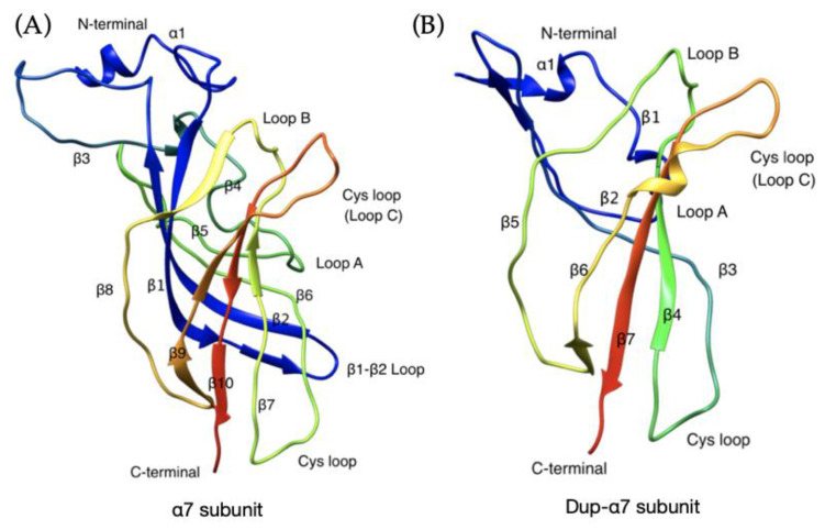
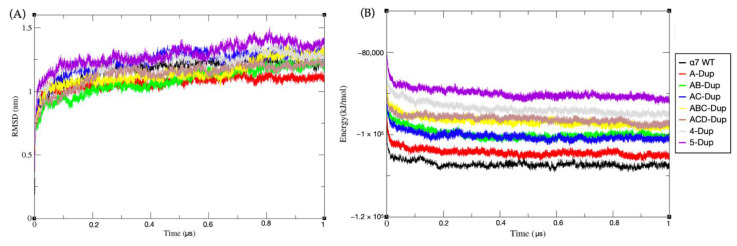
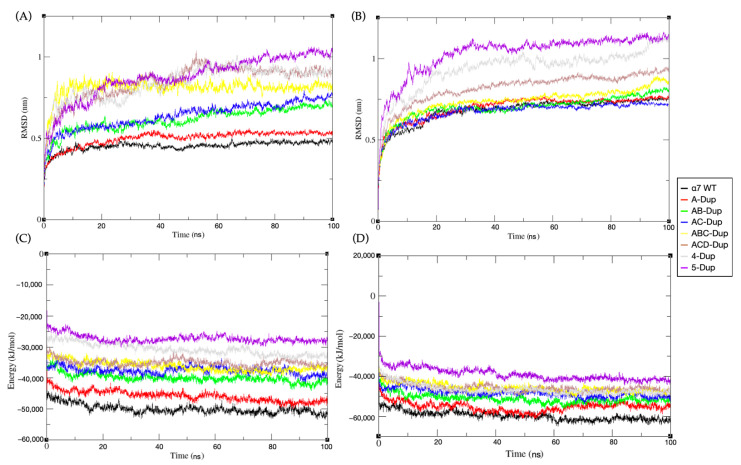
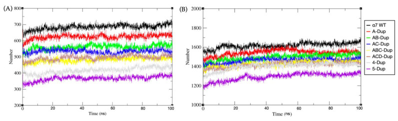
Cholinergic α7 nicotinic receptors encoded by the CHRNA7 gene are ligand-gated ion channels directly related to memory and immunomodulation. Exons 5-7 in CHRNA7 can be duplicated and fused to exons A-E of FAR7a, resulting in a hybrid gene known as CHRFAM7A, unique to humans. Its product, denoted herein as Dupα7, is a truncated subunit where the N-terminal 146 residues of the ligand binding domain of the α7 receptor have been replaced by 27 residues from FAM7. Dupα7 negatively affects the functioning of α7 receptors associated with neurological disorders, including Alzheimer's diseases and schizophrenia. However, the stoichiometry for the α7 nicotinic receptor containing dupα7 monomers remains unknown. In this work, we developed computational models of all possible combinations of wild-type α7 and dupα7 pentamers and evaluated their stability via atomistic molecular dynamics and coarse-grain simulations. We assessed the effect of dupα7 subunits on the Ca2+ conductance using free energy calculations. We showed that receptors comprising of four or more dupα7 subunits are not stable enough to constitute a functional ion channel. We also showed that models with dupα7/α7 interfaces are more stable and are less detrimental for the ion conductance in comparison to dupα7/dupα7 interfaces. Based on these models, we used protein-protein docking to evaluate how such interfaces would interact with an antagonist, α-bungarotoxin, and amyloid Aβ42. Our findings show that the optimal stoichiometry of dupα7/α7 functional pentamers should be no more than three dupα7 monomers, in favour of a dupα7/α7 interface in comparison to a homodimer dupα7/dupα7 interface. We also showed that receptors bearing dupα7 subunits are less sensitive to Aβ42 effects, which may shed light on the translational gap reported for strategies focused on nicotinic receptors in 'Alzheimer's disease research.
Keywords: CHRFAM7A; CHRNA7; coarse grain simulation; molecular dynamics; umbrella simulations; α7 nicotinic receptors.
Conflict of interest statement
The authors declare no conflict of interest.
Figures
Similar articles
-
The Human-Restricted Isoform of the α7 nAChR, CHRFAM7A: A Double-Edged Sword in Neurological and Inflammatory Disorders.Int J Mol Sci. 2022 Mar 22;23(7):3463. doi: 10.3390/ijms23073463. Int J Mol Sci. 2022. PMID: 35408823 Free PMC article. Review.
-
A human-specific, truncated α7 nicotinic receptor subunit assembles with full-length α7 and forms functional receptors with different stoichiometries.J Biol Chem. 2018 Jul 6;293(27):10707-10717. doi: 10.1074/jbc.RA117.001698. Epub 2018 May 21. J Biol Chem. 2018. PMID: 29784875 Free PMC article.
-
The human-specific duplicated α7 gene inhibits the ancestral α7, negatively regulating nicotinic acetylcholine receptor-mediated transmitter release.J Biol Chem. 2021 Jan-Jun;296:100341. doi: 10.1016/j.jbc.2021.100341. Epub 2021 Jan 28. J Biol Chem. 2021. PMID: 33515545 Free PMC article.
-
The duplicated α7 subunits assemble and form functional nicotinic receptors with the full-length α7.J Biol Chem. 2014 Sep 19;289(38):26451-26463. doi: 10.1074/jbc.M114.582858. Epub 2014 Jul 23. J Biol Chem. 2014. PMID: 25056953 Free PMC article.
-
The human CHRNA7 and CHRFAM7A genes: A review of the genetics, regulation, and function.Neuropharmacology. 2015 Sep;96(Pt B):274-88. doi: 10.1016/j.neuropharm.2015.02.006. Epub 2015 Feb 19. Neuropharmacology. 2015. PMID: 25701707 Free PMC article. Review.
Cited by
-
The Human-Restricted Isoform of the α7 nAChR, CHRFAM7A: A Double-Edged Sword in Neurological and Inflammatory Disorders.Int J Mol Sci. 2022 Mar 22;23(7):3463. doi: 10.3390/ijms23073463. Int J Mol Sci. 2022. PMID: 35408823 Free PMC article. Review.
-
Sentinel Lymph Node Gene Expression Signature Predicts Recurrence-Free Survival in Cutaneous Melanoma.Cancers (Basel). 2022 Oct 11;14(20):4973. doi: 10.3390/cancers14204973. Cancers (Basel). 2022. PMID: 36291758 Free PMC article.
References
-
- Maroli A., Di Lascio S., Drufuca L., Cardani S., Setten E., Locati M., Fornasari D., Benfante R. Effect of donepezil on the expression and responsiveness to LPS of CHRNA7 and CHRFAM7A in macrophages: A possible link to the cholinergic anti-inflammatory pathway. J. Neuroimmunol. 2019;332:155–166. doi: 10.1016/j.jneuroim.2019.04.012. - DOI - PubMed
MeSH terms
Substances
Grants and funding
LinkOut - more resources
Full Text Sources
Medical
Miscellaneous
