. 2021 May 22;4(2):38.
doi: 10.3390/mps4020038.

Conformational Control of DNA Origami by DNA Oligomers, Intercalators and UV Light

Affiliations

Conformational Control of DNA Origami by DNA Oligomers, Intercalators and UV Light

Ruixin Li et al. Methods Protoc. .

Abstract

DNA origami has garnered great attention due to its excellent programmability and precision. It offers a powerful means to create complex nanostructures which may not be possible by other methods. The macromolecular structures may be used as static templates for arranging proteins and other molecules. They are also capable of undergoing structural transformation in response to external signals, which may be exploited for sensing and actuation at the nanoscale. Such on-demand reconfigurations are executed mostly by DNA oligomers through base-pairing and/or strand displacement, demonstrating drastic shape changes between two different states, for example, open and close. Recent studies have developed new mechanisms to modulate the origami conformation in a controllable, progressive manner. Here we present several methods for conformational control of DNA origami nanostructures including chemical adducts and UV light as well as widely applied DNA oligomers. The detailed methods should be useful for beginners in the field of DNA nanotechnology.

Keywords: DNA origami; UV; conformation; intercalator; strand displacement; stress.


Conflict of interest statement

The authors declare no conflict of interest.

Figures

Figure 1
Figure 1
Schematics of the conformational change in a DNA origami. (a) Layout of scaffold pattern in a rectangular origami tile. (b) A section of the origami tile indicating the periodic arrangement of the staples (blue and gray) against the scaffold (black). (c) The repeating segment indicated as a red dashed box in (b). The staples (blue and gray) pair with the scaffold (black), forming double helices connected by crossovers (indicated by blue dotted circles). There are 32 bp between the adjacent crossovers for three turns, corresponding to 10.67 bp/turn. (df) Side views of the periodic arrangement in (c) along with corresponding origami conformations from finite element simulations using CanDo on the right. (d) The DNA duplexes experience significant torsional deformation under natural conditions (10.5 bp/turn) in order to maintain crossover connections. The helical mismatch results in a right-handed twist over the entire origami tile. If the mismatch is compensated, crossovers will form without distortion. As such, the tile will be perfectly flat (e). Over-compensating the mismatch (e.g., 10.84 bp/turn) will lead to a left-handed twist in the origami tile (f). (Contents in this figure were previously published. Adapted with permission from [42]. Copyright (2016) American Chemical Society. Further permissions related to the material excerpted should be directed to the ACS).
Figure 2
Figure 2
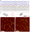
(a) Schematic of a portion of a single-layer DNA origami rectangle with its upper and lower boundaries (black and blue) connected and sealed by a set of linker strands (red) with toeholds (green) at both ends. Staple strands in the origami tile are shown in gray. (b) Schematic of DNA origami cyclization. A flat tile bends and cyclizes into a short cylinder with the help of the linkers (red). Gray rods represent DNA double helices. The sealed boundaries are indicated by red lines. (c,d) AFM images of the two origami tile species. (c) All origami tiles formed by thermal annealing are flat. (d) Cyclized DNA origami after incubation of flat tiles with linkers at 50 °C for 2 h. The majority of DNA origami measures half the length of the flat tile with twice thickness. (Contents in this figure were previously published. Adapted with permission from [37]. Copyright (2014) American Chemical Society).
Figure 3
Figure 3
(ae) AFM images of DNA origami ribbons (2 nM) as a function of EtBr concentration from 0 to 3.5 µM. CanDo simulation results for 10.5 to 10.8 bp/turn are also shown. The handedness of the ribbon twist is represented by the shape of the parallelogram kinks highlighted in (b,d). The added EtBr intercalates into DNA duplexes, unwinding the DNA helicity and increasing the helical pitch. As a result, the ribbon conformation gradually changes from a right-handed twist to flat and then to a left-handed curvature. The progressive conformational change is characterized by the kink density. (f,g) Statistics of kink density in the ribbons. (f) Histogram of kink-kink distance measured under two EtBr concentrations: 0 and 2 µM. In the inset, the parallelogram-shaped kinks indicating the degree of origami twisting are marked using software ImageJ. Distances between the right-handed and left-handed kinks are marked as positive and negative, respectively. The statistics follow Gaussian functions in general. (g) Kink density, defined as the inverse of the distance between neighboring kinks, decreases as EtBr amount increases. The plot suggests that flattening is expected to occur at approximately 1 µM EtBr, which agrees well with experiment. The inverse of the average distance and the associated standard deviation are denoted by red symbols. The black symbols indicate the scattering of the measurements. The blue line suggests the progressive control of origami conformation using intercalators. (Contents in this figure were previously published. Adapted with permission from [42]. Copyright (2016) American Chemical Society. Further permissions related to the material excerpted should be directed to the ACS).
Figure 4
Figure 4
Effect of UV light on DNA structures. (a) Schematic of an origami rectangle. The tile is designed at 10.67 bp/turn, and the internal stress causes a global curvature. Moderate UVC or UVB radiation flattens the rectangular origami tile. (b) AFM image of polymerized DNA origami ribbons. The curvature of the individual tiles leads to a heavy twist of the ribbons. The parallelogram-shaped kinks indicate the right-handed twist (also see Figure 3). (c) AFM image of DNA ribbons after UVC irradiation (~2.5 kJ/m2). The origami structures are completely flat. (Contents in this figure were previously published. Adapted with permission from [60]. Copyright (2017) American Chemical Society. Further permissions related to the material excerpted should be directed to the ACS).
Figure 5
Figure 5
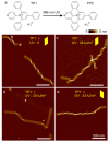
(a) Chemical structure of photoswitchable intercalator: TP1 and TP2. TP1 does not associate with DNA. UVA converts TP1 to TP2, which is a strong DNA intercalator. (be) AFM images of origami ribbons with and without TP1 and UVA radiation. (b,c) If one of the two components is absent, there is no change in the origami structure. (d,e) Conformation change occurs only when both TP1 and UVA are present simultaneously. (d) Partial conversion of TP1 to TP2 flattens DNA structures. (e) Complete conversion can change the DNA helicity significantly, causing a flip of twist handedness. Yellow parallelograms highlight the shape of the kinks, suggesting twist-handedness. (Contents in this figure were previously published. Adapted with permission from [60]. Copyright (2017) American Chemical Society. Further permissions related to the material excerpted should be directed to the ACS).

Similar articles

Cited by

References

    1. Seeman N.C. DNA in a Material World. Nature. 2003;421:427–431. doi: 10.1038/nature01406. - DOI - PubMed
    1. Jin Z., Sun W., Ke Y., Shih C.-J., Paulus G.L.C., Wang Q.H., Mu B., Yin P., Strano M.S. Metallized DNA Nanolithography for Encoding and Transferring Spatial Information for Graphene Patterning. Nat. Commun. 2013;4:1663. doi: 10.1038/ncomms2690. - DOI - PubMed
    1. Pinheiro A.V., Han D., Shih W.M., Yan H. Challenges and Opportunities for Structural DNA Nanotechnology. Nat. Nanotechnol. 2011;6:763–772. doi: 10.1038/nnano.2011.187. - DOI - PMC - PubMed
    1. Sobczak J.-P.J., Martin T.G., Gerling T., Dietz H. Rapid Folding of DNA into Nanoscale Shapes at Constant Temperature. Science. 2012;338:1458–1461. doi: 10.1126/science.1229919. - DOI - PubMed
    1. Han D., Pal S., Nangreave J., Deng Z., Liu Y., Yan H. DNA Origami with Complex Curvatures in Three-Dimensional Space. Science. 2011;332:342–346. doi: 10.1126/science.1202998. - DOI - PubMed

LinkOut - more resources