. 2021 May 22;10(6):1288.
doi: 10.3390/cells10061288.

Generators of Pressure-Evoked Currents in Vertebrate Outer Retinal Neurons

Affiliations

Generators of Pressure-Evoked Currents in Vertebrate Outer Retinal Neurons

Ji-Jie Pang et al. Cells. .

Abstract

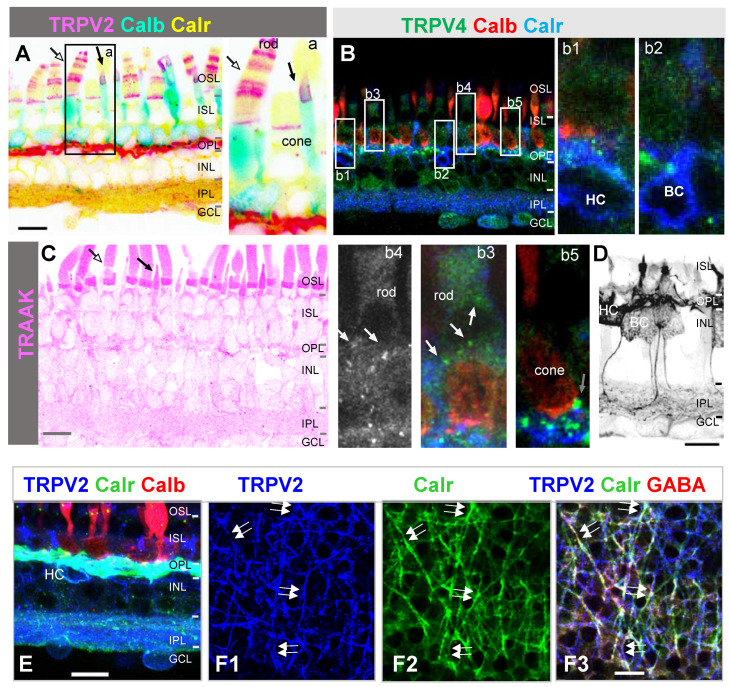
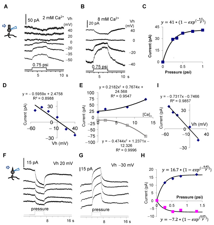
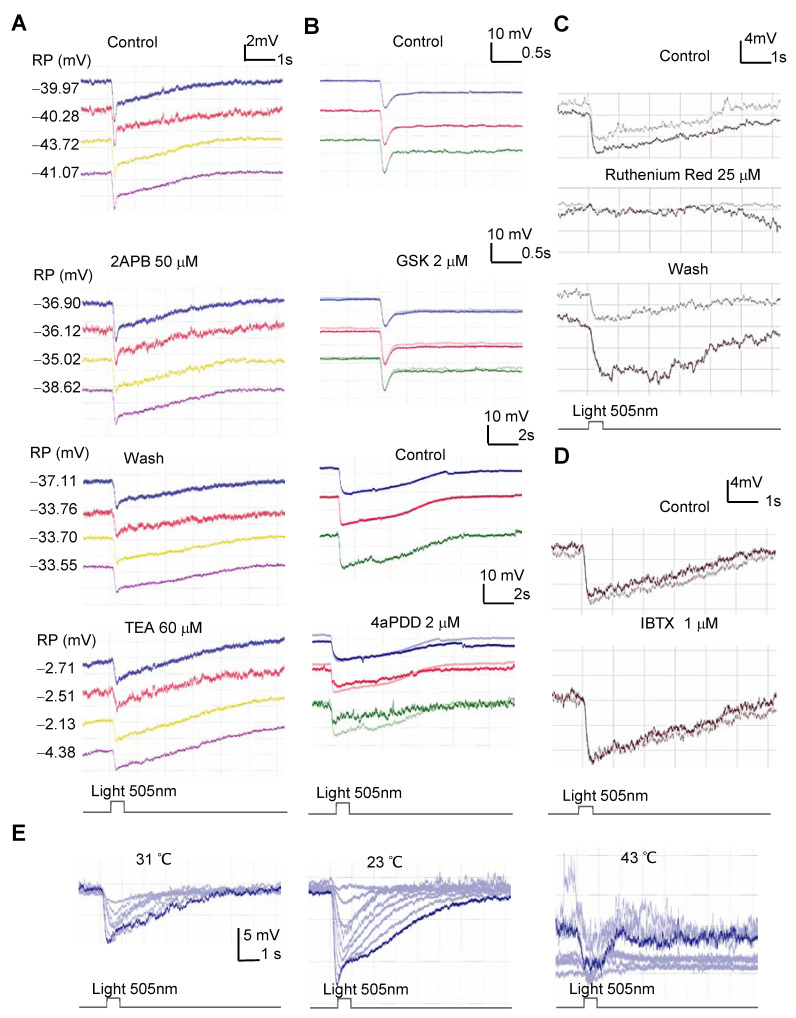
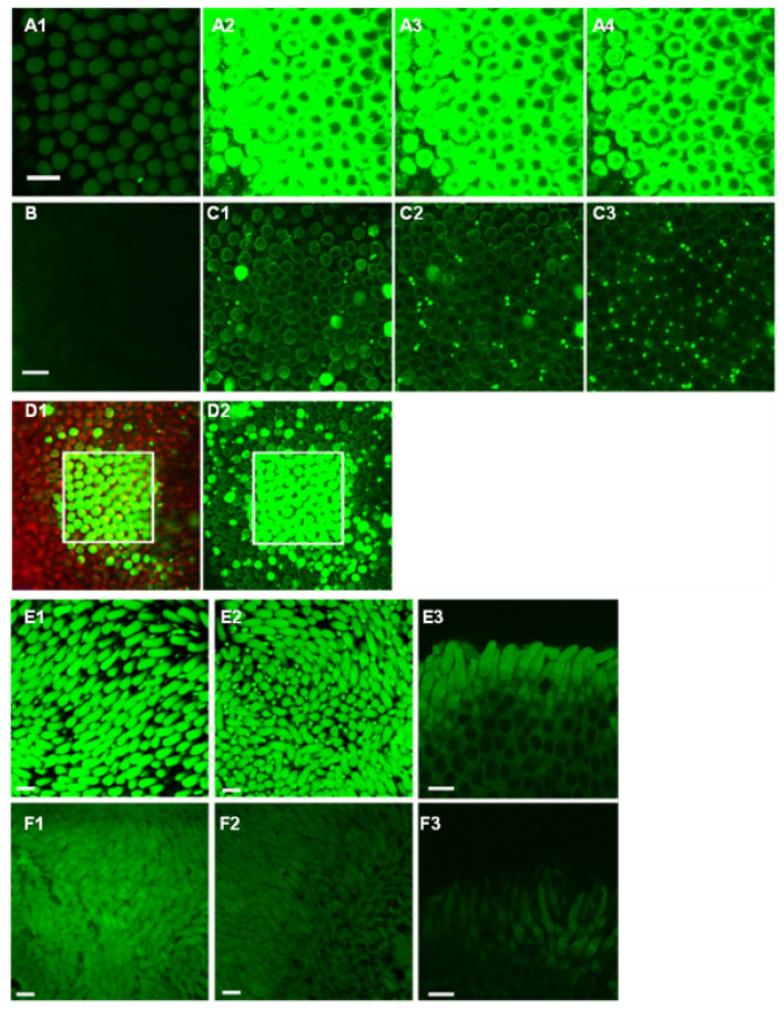
(1) Background: High-tension glaucoma damages the peripheral vision dominated by rods. How mechanosensitive channels (MSCs) in the outer retina mediate pressure responses is unclear. (2) Methods: Immunocytochemistry, patch clamp, and channel fluorescence were used to study MSCs in salamander photoreceptors. (3) Results: Immunoreactivity of transient receptor potential channel vanilloid 4 (TRPV4) was revealed in the outer plexiform layer, K+ channel TRAAK in the photoreceptor outer segment (OS), and TRPV2 in some rod OS disks. Pressure on the rod inner segment evoked sustained currents of three components: (A) the inward current at <-50 mV (Ipi), sensitive to Co2+; (B) leak outward current at ≥-80 mV (Ipo), sensitive to intracellular Cs+ and ruthenium red; and (C) cation current reversed at ~10 mV (Ipc). Hypotonicity induced slow currents like Ipc. Environmental pressure and light increased the FM 1-43-identified open MSCs in the OS membrane, while pressure on the OS with internal Cs+ closed a Ca2+-dependent current reversed at ~0 mV. Rod photocurrents were thermosensitive and affected by MSC blockers. (4) Conclusions: Rods possess depolarizing (TRPV) and hyperpolarizing (K+) MSCs, which mediate mutually compensating currents between -50 mV and 10 mV, serve as an electrical cushion to minimize the impact of ocular mechanical stress.

Keywords: TRPV; confocal microscopy; immunofluorescence; mechanosensitive channel; patch clamping; potassium channel; rod; temperature.


Conflict of interest statement

The authors declare no conflict of interests.

Figures

Figure 1
Figure 1
The expression of several MSCs in the salamander retina. Confocal images show the retina slice triple-labeled for calbindin D-28k (Calb), calretinin (Calr), and TRPV2 (A,E,F) or TRPV4 (B) or probed for TRAAK (C). (A) TRPV2 antibody brightly labels the outer plexiform layer (OPL), half disks in the rod outer segment (OS) (white arrow), and cone OS (black arrow) and weakly reveals some processes in the inner plexiform layer (IPL) and somas in the ganglion cell layer (GCL). (B) Bright TRPV4-immunoreactive puncta are mostly present in the OPL and terminals of cones (black arrow, (b5)), while weaker TRPV4 signals are visible in somas and dendrites of horizontal cells (HCs, (b1)), bipolar cells (BCs) (b2), the basal membrane of rods and putative rod axon terminals (white arrow, (b3,b4)), the inner plexiform layer (IPL), and somas in the GCL. (C) TRAAK antibody labeled the OS of single cones (black arrow) the most brightly, and it clearly revealed the OS of rods (white arrow). Weaker TRAAK signals are sparsely present in the OPL and IPL. (D) Calretinin antibody heavily labeled HCs and clearly labeled some BCs. (E) Calretinin-labeled soma of HCs positive for TRPV2. (F) At the OPL focal plane, nearly half dendrites of HCs that are identified by calretinin (F2) and GABA ((F3), red) are labeled for TRPV2 (F1) (white double-arrow). (F1,F2) display the blue and green channels of (F3), respectively. OSL: outer segment layer; ISL: inner segment layer; RGC: retinal ganglion cell. Scale bars: 20 µm.
Figure 2
Figure 2
Pressure-evoked currents in rods. Rods are recorded in voltage-clamp mode with a patch pipette filled with an internal solution containing Cs+ 137 mM (B) or K+ 132 mM (C) and labeled with Lucifer yellow (A). (B) A rod responds to light steps with outward currents (B1) at different holding potentials (Vh), and the short dynamic pressure pulses applied with a puffer pipette to the soma of the rod elicited primarily inward currents (Ipi) (B2). Ruthenium red (RR) shifted the reversal potential from −50 mV to ~ −20 mV and unmasked a non-selective cation conductance (Ipc) (B3). (C1) Longer pressure steps were applied to the outer plexiform layer and evoked currents reversed around −50 mV. (C2) In the presence of synaptic blocker Co2+, the evoked current was primarily outward (Ipo). (C3) In the presence of Co2+ and RR, the evoked current reversed at ~10 mV, unmasking a non-selective cation conductance (Ipc). (C4) The outward current was washed back. (C5) In a hypotonic bath solution, a hypertonic puffer solution was applied with a dynamic pressure step and induced a slow decrease in the non-selective cation conductance (triangle), which was reversed around −20 mV and superimposed on an outward current (dot). (C6) The same dynamic pressure from the same puffer pipette caused a rather small outward current on a clean patch pipette placed at the same distance. (D) The response of a rod to both light and pressure recorded with a Cs+-containing internal solution. The results demonstrate three major components of the pressure-evoked current in rods.
Figure 3
Figure 3
Response of photoreceptors to pressure applied to the outer segment. (A,B,F,G) depict the pressure-evoked current. With the internal Cs+ blockage of K+ channels, the pressure causes a sustained decrease in the cation conductance in a rod (A,B), which reverses at ~5 mV ((D), at 0.5 psi) and saturates at ~0.5 psi (C). (E) The amplitude of the pressure-evoked current at −30 mV (diamond) and 20 mV (square) increases upon increasing the extracellular Ca2+ concentration ([Ca2+]o) (two-tailed p = 0.0229 and 0.0002, respectively). The pressure–current response curve was well fit to an exponential rise to the maximum function. The two-tailed p for Rmax is 0.0001, and p for b is 0.0004. F and G show the pressure-evoked current in a cone at −30 mV (G) and 20 mV (F), which reverses near 0 mV (I), and the pressure–current response curve (H) was well fit to two exponential functions. For the upper equation (−30 mV), the two-tailed p for Rmax and b is 0.0001 and 0.0020, respectively. For the lower equation (20 mV), the two-tailed p for Rmax and b is 0.0080 and 0.0442, respectively. The data indicate that the extracellular pressure applied to the outer segment causes the closure of TRPV2 in the outer segment of photoreceptors.
Figure 4
Figure 4
The effect of modulators of the mechanical sensitive ion channels on the light response of rods. Rods were recorded for the light response in current-clamp mode. (A,B) Three to four rods were simultaneously recorded with the multi-cell patch-clamp technique. TRPV2 agonist 2APB weakly depolarizes rods and slightly reduces the light response (A). The K+ channel blocker TEA largely depolarizes rods and reduces the light response. (B) TRPV4 channel agonists GSK and 4aPDD slightly hyperpolarize rods (the highlighted trace). (C) The light response in a rod was largely and reversibly reduced by ruthenium red (RR). (D) A blocker for BK channels, IBTX, enhances the light response of a rod. The two traces in each panel in C and D were recorded 2–3 min apart. (E) The light-evoked potentials recorded at eight light intensities are larger at 23 °C than at 31 °C and severely disrupted at 43 °C. (BE) Membrane potentials were maintained at ~−40 mV.
Figure 5
Figure 5
The modification of light (AD) and pressure (E,F) (28 mmHg, (E)) on the FM 1-43-labeled open channels. (A) A selected area during the incubation of FM 1-43 at the focal plane of the outer segment (OS). (A1A4) were taken from 0 s, 20 s, 50 s (begins wash), and 2 min after applying the drug, respectively (B) The OPL of the selected area after 20 min. (C) Other areas at the OS plane after 49 min, showing a low density of labeled rods (C1) and single and double cones (C2,C3). Labeled double cones are less restricted to the selected region. All images in A to C were taken under the same conditions. (D) A large region including the selected area (white square) after 66 min. The green channel in (D1,D2) shows FM 1-43, and the red channel in D1 is the Nomarski image. The data indicate that the 488 nm laser illumination opened some channels permeable to FM 1-43. (E) A piece of the flat-mount retina was simultaneously exposed to 3 µM FM 1-43 plus 28 mmHg for 80 s compared with the other piece from the same eye that was exposed to the same drug without the pressure (F). (E1E3) and (F1F3) are images from different locations. (E3,F3) were taken from the edge of the tissue. The fluorescence is the brightest in the outer segment and enhanced by the pressure. All images in (E,F) were taken under the same conditions. Scale bars: 20 µm. The data indicate that light and pressure open channels permeable to FM 1-43.

Similar articles

Cited by

References

    1. Quigley H.A. Glaucoma. Lancet. 2011;377:1367–1377. doi: 10.1016/S0140-6736(10)61423-7. - DOI - PubMed
    1. Harasymowycz P., Birt C., Gooi P., Heckler L., Hutnik C., Jinapriya D., Shuba L., Yan D., Day R. Medical Management of Glaucoma in the 21st Century from a Canadian Perspective. J. Ophthalmol. 2016;2016:6509809. doi: 10.1155/2016/6509809. - DOI - PMC - PubMed
    1. Wostyn P., De Groot V., Audenaert K., De Deyn P.P. Are intracranial pressure fluctuations important in glaucoma? Med. Hypotheses. 2011;77:598–600. doi: 10.1016/j.mehy.2011.06.043. - DOI - PubMed
    1. Caprioli J., Coleman A.L. Intraocular pressure fluctuation a risk factor for visual field progression at low intraocular pressures in the advanced glaucoma intervention study. Ophthalmology. 2008;115:1123–1129. doi: 10.1016/j.ophtha.2007.10.031. - DOI - PubMed
    1. Asrani S., Zeimer R., Wilensky J., Gieser D., Vitale S., Lindenmuth K. Large diurnal fluctuations in intraocular pressure are an independent risk factor in patients with glaucoma. J. Glaucoma. 2000;9:134–142. doi: 10.1097/00061198-200004000-00002. - DOI - PubMed

Publication types