

Automatic Detection of Mandibular Fractures in Panoramic Radiographs Using Deep Learning
- PMID: 34067462
- PMCID: PMC8224557
- DOI: 10.3390/diagnostics11060933
Automatic Detection of Mandibular Fractures in Panoramic Radiographs Using Deep Learning
Abstract
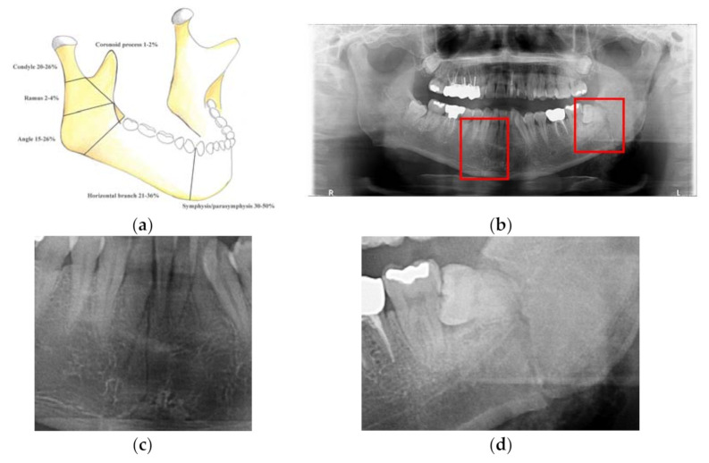
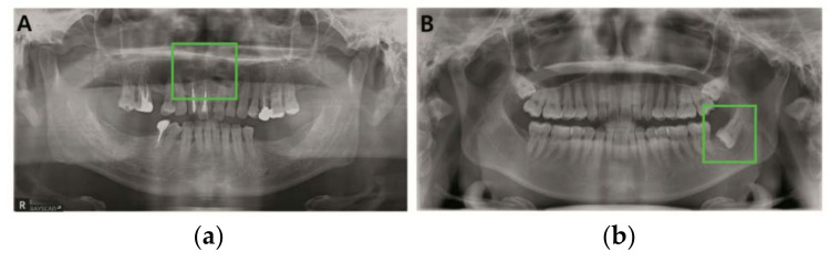
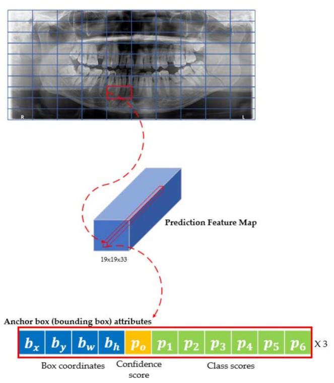
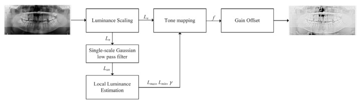
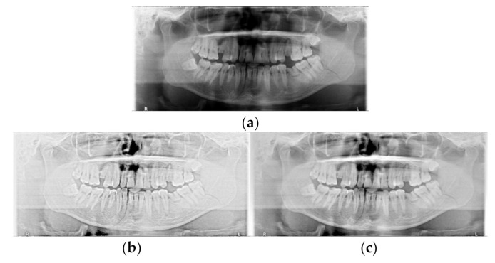
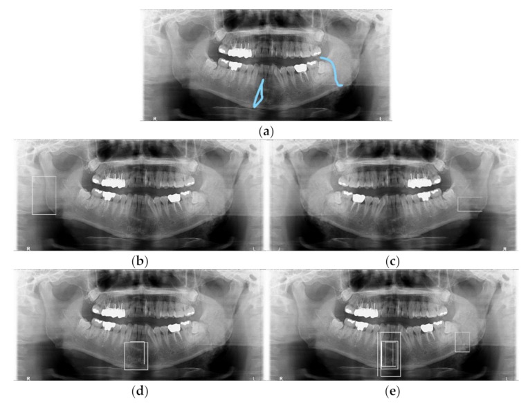
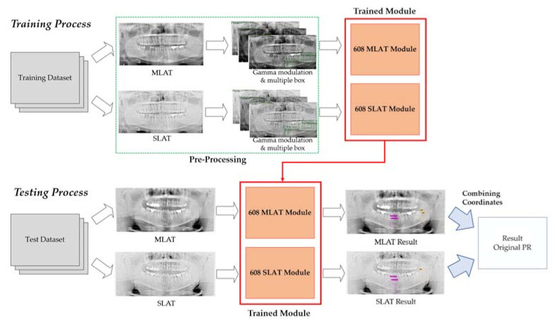
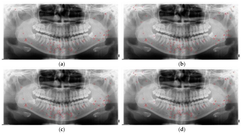
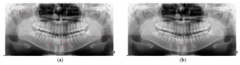
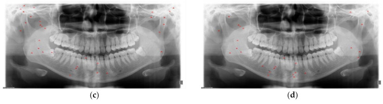
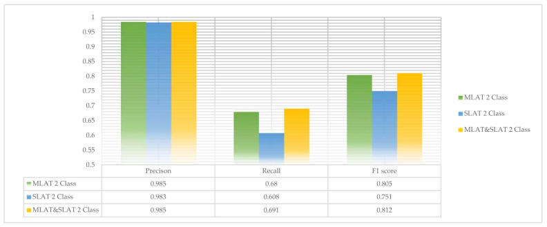
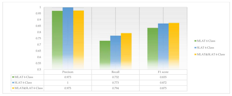
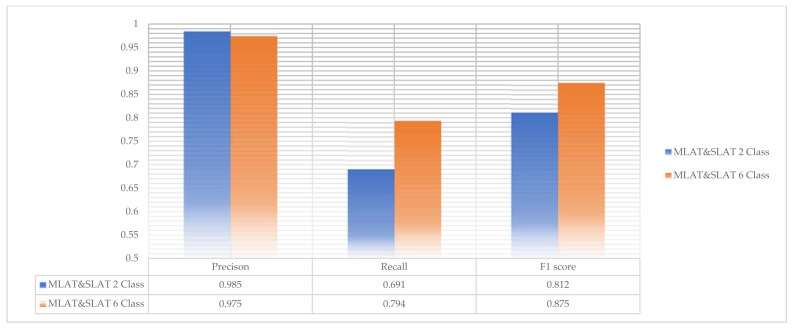
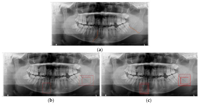
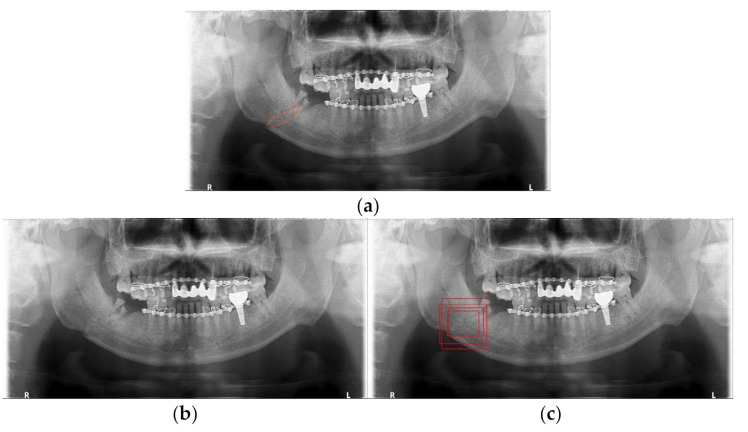
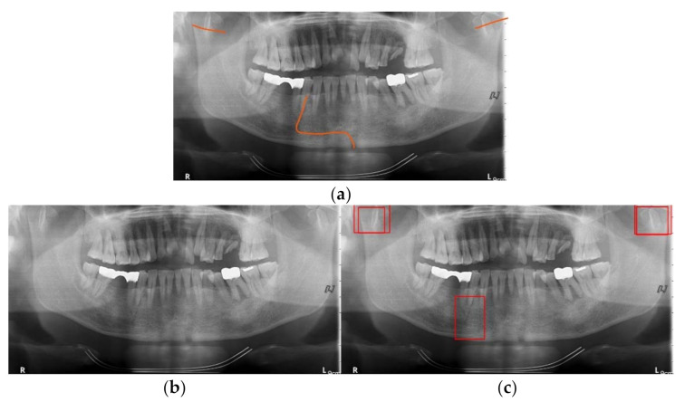
Mandibular fracture is one of the most frequent injuries in oral and maxillo-facial surgery. Radiologists diagnose mandibular fractures using panoramic radiography and cone-beam computed tomography (CBCT). Panoramic radiography is a conventional imaging modality, which is less complicated than CBCT. This paper proposes the diagnosis method of mandibular fractures in a panoramic radiograph based on a deep learning system without the intervention of radiologists. The deep learning system used has a one-stage detection called you only look once (YOLO). To improve detection accuracy, panoramic radiographs as input images are augmented using gamma modulation, multi-bounding boxes, single-scale luminance adaptation transform, and multi-scale luminance adaptation transform methods. Our results showed better detection performance than the conventional method using YOLO-based deep learning. Hence, it will be helpful for radiologists to double-check the diagnosis of mandibular fractures.
Keywords: YOLO; YOLO v4; deep learning; image processing; mandibular fracture; multi-scale luminance adaptation transform (MLAT); object detection; panoramic radiography; single-scale luminance adaptation transform (SLAT).
Conflict of interest statement
The authors declare that there is no conflict of interests regarding the publication of this paper.
Figures
Similar articles
-
Combined Deep Learning Techniques for Mandibular Fracture Diagnosis Assistance.Life (Basel). 2022 Oct 26;12(11):1711. doi: 10.3390/life12111711. Life (Basel). 2022. PMID: 36362866 Free PMC article.
-
Automated permanent tooth detection and numbering on panoramic radiograph using a deep learning approach.Oral Surg Oral Med Oral Pathol Oral Radiol. 2024 May;137(5):537-544. doi: 10.1016/j.oooo.2023.06.003. Epub 2023 Jun 8. Oral Surg Oral Med Oral Pathol Oral Radiol. 2024. PMID: 37633788
-
Assessment of deep convolutional neural network models for mandibular fracture detection in panoramic radiographs.Int J Oral Maxillofac Surg. 2022 Nov;51(11):1488-1494. doi: 10.1016/j.ijom.2022.03.056. Epub 2022 Apr 6. Int J Oral Maxillofac Surg. 2022. PMID: 35397969
-
Deep learning model for the automated evaluation of contact between the lower third molar and inferior alveolar nerve on panoramic radiography.J Dent Sci. 2023 Jul;18(3):991-996. doi: 10.1016/j.jds.2022.12.008. Epub 2022 Dec 26. J Dent Sci. 2023. PMID: 37404620 Free PMC article.
-
A deep-learning artificial intelligence system for assessment of root morphology of the mandibular first molar on panoramic radiography.Dentomaxillofac Radiol. 2019 Mar;48(3):20180218. doi: 10.1259/dmfr.20180218. Epub 2018 Nov 9. Dentomaxillofac Radiol. 2019. PMID: 30379570 Free PMC article.
Cited by
-
Combined Deep Learning Techniques for Mandibular Fracture Diagnosis Assistance.Life (Basel). 2022 Oct 26;12(11):1711. doi: 10.3390/life12111711. Life (Basel). 2022. PMID: 36362866 Free PMC article.
-
Assessing the reliability of CBCT-based AI-generated STL files in diagnosing osseous changes of the mandibular condyle: a comparative study with ground truth diagnosis.Dentomaxillofac Radiol. 2023 Oct;52(7):20230141. doi: 10.1259/dmfr.20230141. Epub 2023 Sep 4. Dentomaxillofac Radiol. 2023. PMID: 37641960 Free PMC article.
-
The Application of Deep Learning on CBCT in Dentistry.Diagnostics (Basel). 2023 Jun 14;13(12):2056. doi: 10.3390/diagnostics13122056. Diagnostics (Basel). 2023. PMID: 37370951 Free PMC article. Review.
-
Evaluation of deep learning and convolutional neural network algorithms for mandibular fracture detection using radiographic images: A systematic review and meta-analysis.Imaging Sci Dent. 2024 Sep;54(3):232-239. doi: 10.5624/isd.20240038. Epub 2024 Aug 12. Imaging Sci Dent. 2024. PMID: 39371302 Free PMC article. Review.
-
Detection of maxillary sinus pathologies using deep learning algorithms.Eur Arch Otorhinolaryngol. 2025 May 20. doi: 10.1007/s00405-025-09451-4. Online ahead of print. Eur Arch Otorhinolaryngol. 2025. PMID: 40394252
References
-
- Lindh C., Petersson A. Radiologic examination for location of the mandibular canal: A comparison between panoramic radiography and conventional tomography. Int. J. Oral Maxillofac. Implants. 1989;4:249–253. - PubMed
Grants and funding
LinkOut - more resources
Full Text Sources
