

From Melanocytes to Melanoma Cells: Characterization of the Malignant Transformation by Four Distinctly Different Melanin Fluorescence Spectra (Review)
- PMID: 34067690
- PMCID: PMC8156265
- DOI: 10.3390/ijms22105265
From Melanocytes to Melanoma Cells: Characterization of the Malignant Transformation by Four Distinctly Different Melanin Fluorescence Spectra (Review)
Abstract
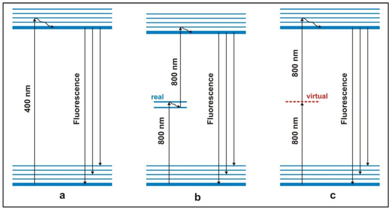
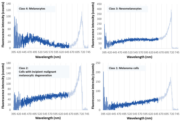
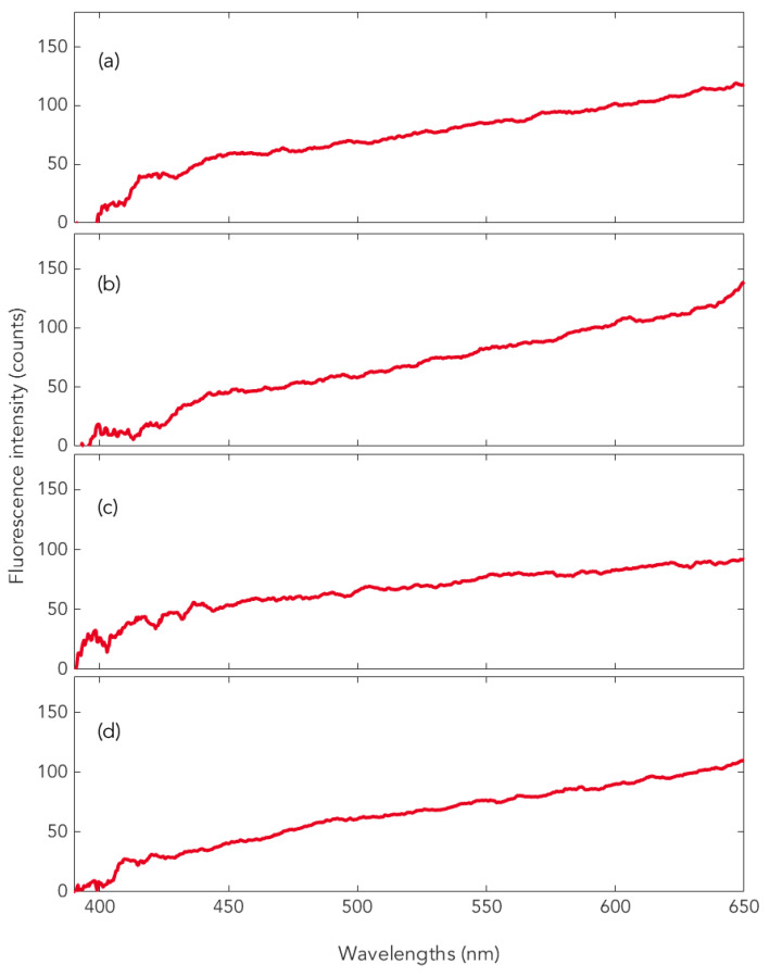
The melanin fluorescence emitted by pigment cells of the human skin has been a central research topic for decades, because melanin, on the one hand, protects against (solar) radiation in the near-UV range, whereas on the other hand, melanocytes are the starting point for the malignant transformation into melanoma. Until recently, however, melanin fluorescence was not accessible in the context of conventional spectroscopy, because it is ultraweak and is overshadowed by the more intense so-called autofluorescence of endogenous fluorophores. The advent of a new method of laser spectroscopy has made this melanin fluorescence measurable in vivo. A stepwise two-photon absorption with 800 nm photons is used, which more selectively excites melanin (dermatofluoroscopy). Our review summarizes the experimental results on melanin fluorescence of the four types of cutaneous pigment cells from healthy and malignant tissues. Outstanding is the finding that different types of melanocytes (i.e., melanocytes of common nevi, versus dysplastic nevi or versus melanoma cells) show characteristically different fluorescence spectra. The possibilities of using this melanin fluorescence for melanoma diagnosis are shown. Moreover, the uniform fluorescence spectra emitted by different melanoma subtypes are essential. Conclusions are drawn about the molecular processes in the melanosomes that determine fluorescence. Finally, experimental suggestions for further investigations are given.
Keywords: dermatofluoroscopy; dysplastic nevi; melanin fluorescence; melanoma subtypes.
Conflict of interest statement
The authors declare no conflict of interest.
Figures
Similar articles
-
The stepwise two-photon excited melanin fluorescence is a unique diagnostic tool for the detection of malignant transformation in melanocytes.Pigment Cell Melanoma Res. 2011 Jun;24(3):438-45. doi: 10.1111/j.1755-148X.2011.00853.x. Epub 2011 May 2. Pigment Cell Melanoma Res. 2011. PMID: 21457482
-
The epidermal melanin unit in the pathophysiology of malignant melanoma.Am J Dermatopathol. 1991 Apr;13(2):179-88. doi: 10.1097/00000372-199104000-00013. Am J Dermatopathol. 1991. PMID: 2029092 Review.
-
Melanin absorption spectroscopy: new method for noninvasive skin investigation and melanoma detection.J Biomed Opt. 2008 Jan-Feb;13(1):014017. doi: 10.1117/1.2844710. J Biomed Opt. 2008. PMID: 18315375
-
Early diagnosis of melanotic melanoma based on laser-induced melanin fluorescence.J Biomed Opt. 2009 May-Jun;14(3):034033. doi: 10.1117/1.3155511. J Biomed Opt. 2009. PMID: 19566326
-
Cutaneous photobiology. The melanocyte vs. the sun: who will win the final round?Pigment Cell Res. 2003 Oct;16(5):434-47. doi: 10.1034/j.1600-0749.2003.00088.x. Pigment Cell Res. 2003. PMID: 12950718 Review.
Cited by
-
Etiologies of Melanoma Development and Prevention Measures: A Review of the Current Evidence.Cancers (Basel). 2021 Sep 30;13(19):4914. doi: 10.3390/cancers13194914. Cancers (Basel). 2021. PMID: 34638397 Free PMC article. Review.
-
Alginate Nanoparticles Containing Cuminum cyminum and Zataria multiflora Essential Oils with Promising Anticancer and Antibacterial Effects.Int J Biomater. 2024 May 2;2024:5556838. doi: 10.1155/2024/5556838. eCollection 2024. Int J Biomater. 2024. PMID: 38725434 Free PMC article.
-
Ornidazole suppresses CD133+ melanoma stem cells via inhibiting hedgehog signaling pathway and inducing multiple death pathways in a mouse model.Croat Med J. 2022 Oct 31;63(5):461-474. doi: 10.3325/cmj.2022.63.461. Croat Med J. 2022. PMID: 36325671 Free PMC article.
-
New Aspects Regarding the Fluorescence Spectra of Melanin and Neuromelanin in Pigmented Human Tissue Concerning Hypoxia.Int J Mol Sci. 2024 Aug 2;25(15):8457. doi: 10.3390/ijms25158457. Int J Mol Sci. 2024. PMID: 39126026 Free PMC article.
-
Immunotherapy as a Turning Point in the Treatment of Melanoma Brain Metastases.Discoveries (Craiova). 2023 Jun 30;11(2):e169. doi: 10.15190/d.2023.8. eCollection 2023 Apr-Jun. Discoveries (Craiova). 2023. PMID: 37583899 Free PMC article. Review.
References
-
- Elder D.E., Bastian B.C., Cree I.A., Massi D., Scolyer R.A. The 2018 World Health Organization classification of cutaneous, mucosal, and uveal melanoma: Detailed analysis of 9 distinct subtypes defined by their evolutionary pathway. Arch. Pathol. Lab. Med. 2020;144:500–522. doi: 10.5858/arpa.2019-0561-RA. - DOI - PubMed
-
- Gallas J.M., Eisner M. Fluorescence of melanin–dependence upon excitation wavelenght and concentration. Photochem. Photobiol. 1987;45:595–600. doi: 10.1111/j.1751-1097.1987.tb07385.x. - DOI
Publication types
MeSH terms
Substances
LinkOut - more resources
Full Text Sources
Medical
