

Design and Numerical Analysis of a Graphene-Coated SPR Biosensor for Rapid Detection of the Novel Coronavirus
- PMID: 34067769
- PMCID: PMC8156410
- DOI: 10.3390/s21103491
Design and Numerical Analysis of a Graphene-Coated SPR Biosensor for Rapid Detection of the Novel Coronavirus
Abstract
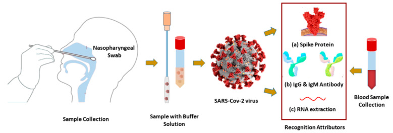
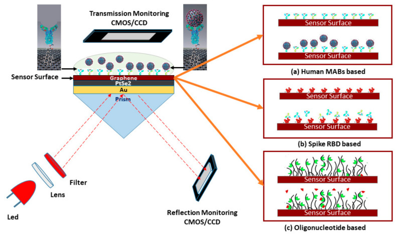
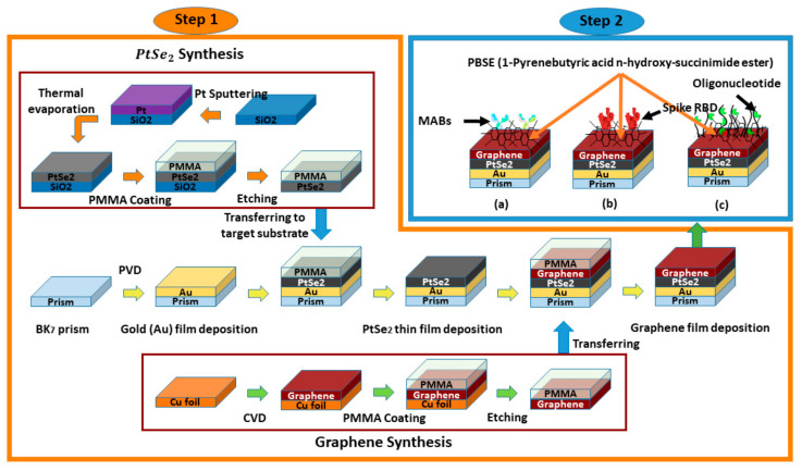
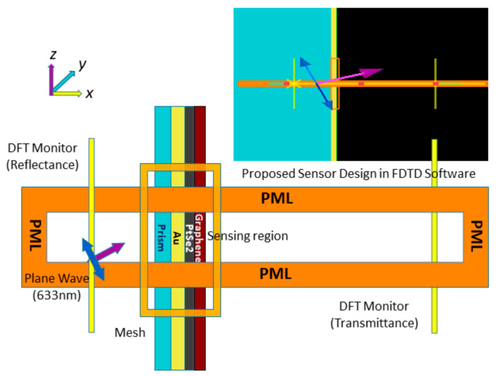
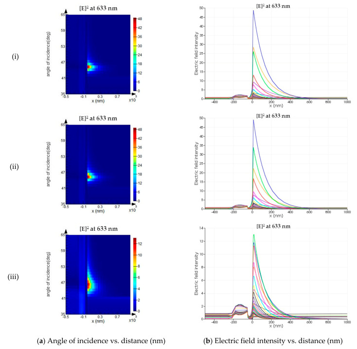
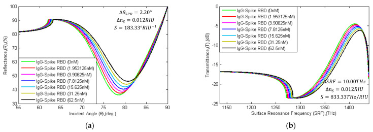
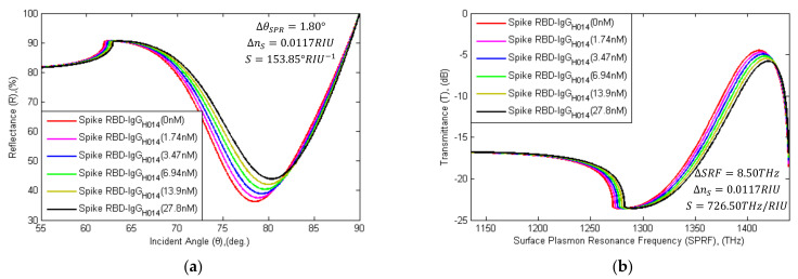
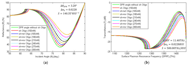
In this paper, a highly sensitive graphene-based multiple-layer (BK7/Au/PtSe2/Graphene) coated surface plasmon resonance (SPR) biosensor is proposed for the rapid detection of the novel Coronavirus (COVID-19). The proposed sensor was modeled on the basis of the total internal reflection (TIR) technique for real-time detection of ligand-analyte immobilization in the sensing region. The refractive index (RI) of the sensing region is changed due to the interaction of different concentrations of the ligand-analyte, thus impacting surface plasmon polaritons (SPPs) excitation of the multi-layer sensor interface. The performance of the proposed sensor was numerically investigated by using the transfer matrix method (TMM) and the finite-difference time-domain (FDTD) method. The proposed SPR biosensor provides fast and accurate early-stage diagnosis of the COVID-19 virus, which is crucial in limiting the spread of the pandemic. In addition, the performance of the proposed sensor was investigated numerically with different ligand-analytes: (i) the monoclonal antibodies (mAbs) as ligand and the COVID-19 virus spike receptor-binding domain (RBD) as analyte, (ii) the virus spike RBD as ligand and the virus anti-spike protein (IgM, IgG) as analyte and (iii) the specific probe as ligand and the COVID-19 virus single-standard ribonucleic acid (RNA) as analyte. After the investigation, the sensitivity of the proposed sensor was found to provide 183.33°/refractive index unit (RIU) in SPR angle (θSPR) and 833.33THz/RIU in SPR frequency (SPRF) for detection of the COVID-19 virus spike RBD; the sensitivity obtained 153.85°/RIU in SPR angle and 726.50THz/RIU in SPRF for detection of the anti-spike protein, and finally, the sensitivity obtained 140.35°/RIU in SPR angle and 500THz/RIU in SPRF for detection of viral RNA. It was observed that whole virus spike RBD detection sensitivity is higher than that of the other two detection processes. Highly sensitive two-dimensional (2D) materials were used to achieve significant enhancement in the Goos-Hänchen (GH) shift detection sensitivity and plasmonic properties of the conventional SPR sensor. The proposed sensor successfully senses the COVID-19 virus and offers additional (1 + 0.55) × L times sensitivity owing to the added graphene layers. Besides, the performance of the proposed sensor was analyzed based on detection accuracy (DA), the figure of merit (FOM), signal-noise ratio (SNR), and quality factor (QF). Based on its performance analysis, it is expected that the proposed sensor may reduce lengthy procedures, false positive results, and clinical costs, compared to traditional sensors. The performance of the proposed sensor model was checked using the TMM algorithm and validated by the FDTD technique.
Keywords: COVID-19; SARS-CoV-2; biosensor; coronavirus; molecular detection; rapid detection; sensor; spike receptor-binding domain; surface plasmon resonance.
Conflict of interest statement
The authors declare no conflict of interest.
Figures
Similar articles
-
Plasmonic photonic biosensor: in situ detection and quantification of SARS-CoV-2 particles.Opt Express. 2022 Oct 24;30(22):40277-40291. doi: 10.1364/OE.469937. Opt Express. 2022. PMID: 36298963
-
A performance comparison of heterostructure surface plasmon resonance biosensor for the diagnosis of novel coronavirus SARS-CoV-2.Opt Quantum Electron. 2023;55(5):448. doi: 10.1007/s11082-023-04700-4. Epub 2023 Mar 25. Opt Quantum Electron. 2023. PMID: 37008732 Free PMC article.
-
Highly Sensitive TiO2/Au/Graphene Layer-Based Surface Plasmon Resonance Biosensor for Cancer Detection.Biosensors (Basel). 2022 Aug 5;12(8):603. doi: 10.3390/bios12080603. Biosensors (Basel). 2022. PMID: 36004999 Free PMC article.
-
An ultra-sensitive surface plasmon resonance biosensor with PtSe2 and BlueP/WS2 heterostructure.Heliyon. 2024 Sep 28;10(19):e38499. doi: 10.1016/j.heliyon.2024.e38499. eCollection 2024 Oct 15. Heliyon. 2024. PMID: 39403496 Free PMC article. Review.
-
Graphene-based nanocomposites for sensitivity enhancement of surface plasmon resonance sensor for biological and chemical sensing: A review.Biosens Bioelectron. 2019 Aug 15;139:111324. doi: 10.1016/j.bios.2019.111324. Epub 2019 May 15. Biosens Bioelectron. 2019. PMID: 31121435 Review.
Cited by
-
Integration of Multiple Interferometers in Highly Multiplexed Diagnostic KITs to Evaluate Several Biomarkers of COVID-19 in Serum.Biosensors (Basel). 2022 Aug 23;12(9):671. doi: 10.3390/bios12090671. Biosensors (Basel). 2022. PMID: 36140055 Free PMC article.
-
Smart City Data Sensing during COVID-19: Public Reaction to Accelerating Digital Transformation.Sensors (Basel). 2021 Jun 8;21(12):3965. doi: 10.3390/s21123965. Sensors (Basel). 2021. PMID: 34201395 Free PMC article.
-
Surface Plasmon Resonance (SPR)- and Localized SPR (LSPR)-Based Virus Sensing Systems: Optical Vibration of Nano- and Micro-Metallic Materials for the Development of Next-Generation Virus Detection Technology.Biosensors (Basel). 2021 Jul 26;11(8):250. doi: 10.3390/bios11080250. Biosensors (Basel). 2021. PMID: 34436053 Free PMC article. Review.
-
Nanomaterial-Based Biosensors for the Detection of COVID-19.Indian J Microbiol. 2025 Mar;65(1):120-136. doi: 10.1007/s12088-024-01336-0. Epub 2024 Jun 23. Indian J Microbiol. 2025. PMID: 40371045
-
Rapid, sensitive and multiplexed detection of SARS-CoV-2 viral nucleic acids enabled by phase-based surface plasmon resonance of metallic gratings.Biomed Opt Express. 2024 Aug 13;15(9):5215-5226. doi: 10.1364/BOE.535051. eCollection 2024 Sep 1. Biomed Opt Express. 2024. PMID: 39296394 Free PMC article.
References
-
- World Health Organization (WHO) Novel Coronavirus 2019 (COVID-19) World Health Organization; Geneva, Switzerland: 2019.
MeSH terms
Substances
LinkOut - more resources
Full Text Sources
Medical
Miscellaneous
