Review
. 2021 May 17;10(5):794.
doi: 10.3390/antiox10050794.

Mitophagy and Oxidative Stress: The Role of Aging

Affiliations
Review

Mitophagy and Oxidative Stress: The Role of Aging

Anna De Gaetano et al. Antioxidants (Basel). .

Abstract

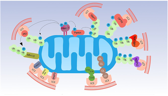
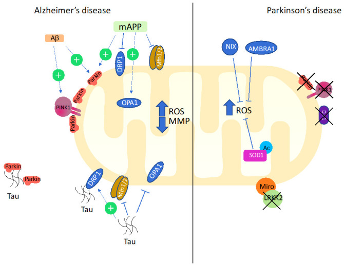
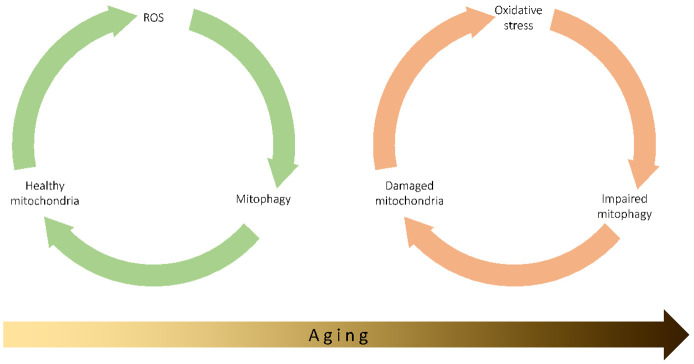
Mitochondrial dysfunction is a hallmark of aging. Dysfunctional mitochondria are recognized and degraded by a selective type of macroautophagy, named mitophagy. One of the main factors contributing to aging is oxidative stress, and one of the early responses to excessive reactive oxygen species (ROS) production is the induction of mitophagy to remove damaged mitochondria. However, mitochondrial damage caused at least in part by chronic oxidative stress can accumulate, and autophagic and mitophagic pathways can become overwhelmed. The imbalance of the delicate equilibrium among mitophagy, ROS production and mitochondrial damage can start, drive, or accelerate the aging process, either in physiological aging, or in pathological age-related conditions, such as Alzheimer's and Parkinson's diseases. It remains to be determined which is the prime mover of this imbalance, i.e., whether it is the mitochondrial damage caused by ROS that initiates the dysregulation of mitophagy, thus activating a vicious circle that leads to the reduced ability to remove damaged mitochondria, or an alteration in the regulation of mitophagy leading to the excessive production of ROS by damaged mitochondria.

Keywords: Alzheimer; PINK1; Parkinson; Reactive Oxygen Species; aging; mitochondria; mitophagy.


Conflict of interest statement

The authors declare no conflict of interest.

Figures

Figure 1
Figure 1
Main molecular mechanisms of mitophagy. Mitophagy is regulated by the interaction of mitochondrial proteins with LC3 through different mechanisms. In the PINK1/Parkin pathway, decreased MMP leads to the accumulation of PINK1 to the OMM. PINK1 phosphorylates both ubiquitin and Parkin. Activated Parkin polyubiquitinates specific proteins on the OMM, making available ubiquitins for PINK1 phosphorylation. The ubiquitinated proteins on the OMM allow the interaction of mitochondria with LC3 through specific adaptors, such as p62, Nbr1, OPTN, TAX1BP1, and NDP52. PINK1 can also phosphorylate Mfn2 and promote its ubiquitination by Parkin and rapid degradation, to prevent fusion of damaged mitochondria with healthy organelles. Besides PINK1/Parkin pathway, mitophagy is triggered by the mitochondrial receptors BNIP3, BNIP3L/NIX, FUNDC1, which can bind directly to LC3. Finally, mitochondrial lipids cardiolipin (CL) and C18-ceramide (C18) can move from the mitochondrial cristae to the OMM, where they interact with LC3.
Figure 2
Figure 2
Main alterations of factors modulating mitophagy observed in AD and PD. In AD, Aβ accumulation upregulated PINK/Parkin pathway. Mutant human APP increases recruitment of Parkin to depolarized mitochondria. mAPP also causes upregulation of mitochondrial fission genes DRP1, and decrease of fusion genes MFN1/2 and OPA1. Tau interacts with Parkin and inhibits its translocation to defective mitochondria. Furthermore, it upregulates fission proteins and inhibits fusion proteins. In PD, defective mitophagy is determined by loss of function of Parkin, PINK1, and DJ- 1; their dysfunction is associated with high levels of ROS. Loss of LRKK2 blocks the degradation of the outer membrane protein Miro and triggers mitophagy. In this context, NIX and AMBRA, can limit the excessive production of ROS. See text for details.
Figure 3
Figure 3
In normal conditions, ROS physiologically produced in the cell, and particularly by mitochondria, can induce mitophagy, which contributes to the normal homeostasis of the cells by removing damaged mitochondria, so maintaining the organelles healthy. The progressive increase in ROS production observed with age can lead to chronic oxidative stress, which in turn impairs mitophagy and reduces the capability to remove damaged mitochondria. Damaged organelles further produced ROS, so keeping a vicious cycle active.

References

    1. Lopez-Otin C., Blasco M.A., Partridge L., Serrano M., Kroemer G. The hallmarks of aging. Cell. 2013;153:1194–1217. doi: 10.1016/j.cell.2013.05.039. - DOI - PMC - PubMed
    1. Yoo S.M., Jung Y.K. A Molecular Approach to Mitophagy and Mitochondrial Dynamics. Mol. Cells. 2018;41:18–26. doi: 10.14348/molcells.2018.2277. - DOI - PMC - PubMed
    1. Vernucci E., Tomino C., Molinari F., Limongi D., Aventaggiato M., Sansone L., Tafani M., Russo M.A. Mitophagy and Oxidative Stress in Cancer and Aging: Focus on Sirtuins and Nanomaterials. Oxidative Med. Cell. Longev. 2019;2019:6387357. doi: 10.1155/2019/6387357. - DOI - PMC - PubMed
    1. Youle R.J., Narendra D.P. Mechanisms of mitophagy. Nat. Rev. Mol. Cell Biol. 2011;12:9–14. doi: 10.1038/nrm3028. - DOI - PMC - PubMed
    1. Ke P.Y. Mitophagy in the Pathogenesis of Liver Diseases. Cells. 2020;9:831. doi: 10.3390/cells9040831. - DOI - PMC - PubMed

LinkOut - more resources