. 2021 May 13;9(5):545.
doi: 10.3390/biomedicines9050545.

Multi-Functional MPT Protein as a Therapeutic Agent against Mycobacterium tuberculosis

Affiliations

Multi-Functional MPT Protein as a Therapeutic Agent against Mycobacterium tuberculosis

Jae-Sung Kim et al. Biomedicines. .

Abstract

Mycobacterium tuberculosis (MTB), the causative agent of tuberculosis (TB), avoids the host immune system through its virulence factors. MPT63 and MPT64 are the virulence factors secreted by MTB which regulate host proteins for the survival and proliferation of MTB in the host. Here, we found that MPT63 bound directly with TBK1 and p47phox, whereas MPT64 interacted with TBK1 and HK2. We constructed a MPT63/64-derived multifunctional recombinant protein (rMPT) that was able to interact with TBK1, p47phox, or HK2. rMPT was shown to regulate IFN-β levels and increase inflammation and concentration of reactive oxygen species (ROS), while targeting macrophages and killing MTB, both in vitro and in vivo. Furthermore, the identification of the role of rMPT against MTB was achieved via vaccination in a mouse model. Taken together, we here present rMPT, which, by regulating important immune signaling systems, can be considered an effective vaccine or therapeutic agent against MTB.

Keywords: MPT peptide; Mycobacterium tuberculosis; TBK1; hexokinase 2; macrophages; p47phox.


Conflict of interest statement

The authors declare that they have no conflict of interest.

Figures

Figure 1
Figure 1
TBK1 and p47phox is bound with MPT63. (A) Identification of TBK1 and p47phox by mass spectrometry analysis in THP-1 cell lysates treated with rMPT63 or rVector. (B) THP-1 cells were stimulated with rMPT63 (5 μg mL−1) for the indicated times, followed by immunoprecipitation (IP) with αHis-agarose bead and IB with αTBK1, αP-TBK1 (S172), αp47phox, αP-p47phox (S304, 345, 359, and 370), αHis, αActin. (C,D) Titration of fluorescently labelled MPT63 with TBK1 and p47phox (left), with Kd (178 and 345 nM) determined by curve fit analysis (right). (E,G) Binding mapping. Schematic diagrams of the structures of MPT63 (upper). At 48 h after transfection with mammalian glutathione S-transferase (GST) or GST-MPT63 and truncated mutant constructs together with Flag-TBK1 or V5-p47phox. 293T cells were used for GST pull down, followed by IB with αFlag or αV5. Cell lysates (WCLs) were used for IB with αFlag or αV5, αGST, and αActin. (F,H) 293T cells are expressing Myc-MPT63 and Flag-TBK1 or V5-p47phox and treated with several Tat-MPT63-N or MPT63-C peptides (10 µM) for 6 h, followed by IP with αMyc and IB with αFlag. WCLs were used for IB with αMyc, αFlag, and αActin. The data are representative of four independent experiments with similar results (AH).
Figure 2
Figure 2
TBK1 and HK2 directly interact with MPT64. (A) Identification of TBK1 and HK2 by mass spectrometry analysis in THP-1 cell lysates treated with rMPT64 or rVector. (B) THP-1 cells were stimulated with rMPT64 (5 μg mL−1) for the indicated times, followed by IP with αHis-agarose bead and IB with αTBK1, αP-TBK1 (S172), αHK2, αHis, αActin. (C,D) Titration of fluorescently labelled MPT64 with TBK1 and HK2 (left), with Kd (193 and 134 nM), determined by curve fit analysis (right). (E,G) Binding mapping. Schematic diagrams of the structures of MPT64 (upper). At 48 h after transfection with GST or GST-MPT63 and truncated mutant constructs together with Flag-TBK1 or V5-p47phox. 293T cells were used for GST pull down, followed by IB with αFlag or αV5. WCLs were used for IB with αFlag or αV5, αGST, and αActin. (F,H) 293T cells expressing Myc-MPT63 and Flag-TBK1 or V5-p47phox and treated with several Tat-MPT64-N or MPT64-C peptides (10 µM) for 6 h, followed by IP with αMyc and IB with αFlag. WCLs were used for IB with αMyc, αFlag, and αActin. The data are representative of four independent experiments with similar results (AH).
Figure 3
Figure 3
TBK1 peptide eliminates MTB by decreasing the STING1–TBK1–IRF3 pathway. (A) Schematic design of TBK1 peptide and its mutants. (B) 293T cells were transfected with V5-IRF3 and/or AU1-STING1 and treated TBK1 peptide or its mutants for 6h (1 µM) with Flag-TBK1 (left) or without Flag-TBK1 (right). 293T cells were used for IP with αFlag or αV5, followed by IB with αV5, αAU1 and αFlag. WCLs were used for IB with αV5, αAU1, αFlag, and αActin. (C,D) THP-1, TBK1+/+ or TBK1−/− BMDM cells were infected by MTB for 4 h and treated TBK1 peptide at various concentrations. After 4 h, THP-1 cells were used for IP with αTBK1 or αIRF3, followed by IB with αIRF3 and αSTING1. WCLs were used for IB with αTBK1, αIRF3, αSTING1, and αActin. (E) TBK1+/+ or TBK1−/− BMDM cells were infected by MTB for 4 h and treated TBK1 peptide at various concentrations. After 18h, supernatants of BMDMs were used for analysis of the level of IFN-β, TNF-α, and IL-6 by ELISA. (F) The burden of MTB was evaluated after 3 d in MTB infected TBK1+/+ or TBK1−/− BMDMs with vehicle or TBK1 peptide. The data are representative of four independent experiments with similar results (BF). Significant differences (* p < 0.05; ** p < 0.01; *** p < 0.001) compared with rVehicle-treated BMDMs.
Figure 4
Figure 4
p47 peptide enhances inflammation against MTB by elevating the level of ROS. (A) Schematic design of p47 peptide (upper). 293T cells were transfected with V5-p47phox and Flag-p22phox or Flag-p67phox and treated p47 peptide for 6 h (1 µM). 293T cells were used for IP with αV5, followed by IB with αFlag. WCLs were used for IB with αFlag, αV5, and αActin. (B) THP-1 or BMDM cells were treated with p47phox in the indicated concentrations. After 18 h, THP-1 or BMDMs cells were used for IP with αp47phox, followed by IB with αp22phox and αp67phox. WCLs were used for IB with αp47phox, αp22phox, αp67phox, αgp91phox, and αActin. (C) p47 peptide mediates the increase of ATP5A1 stability. 293T cells were transfected with V5-p47phox and treated p47 peptide for 24 h. After 24 h, 293T cells were treated with solvent control (SC) or cyclohexamide (CHX, 1 μg mL−1) for the indicated times and cell lysates were used for IB with αV5 and αActin. (D) BMDMs were infected MTB for 4 h and treated Vehicle or p47 peptide of various concentrations for 18 h. To examine the level of ROS, BMDMs measured the fluorescence of DHE, DCFH-DA, mitoSOX to detect O2, H2O2, and mtROS by FACS. (E) BMDMs were infected with MTB for 4 h and treated Vehicle, p47 peptide, or p47 mutant of various concentrations. The supernatant of BMDMs were used for ELISA to measure the level of TNF-α and IL-6. (F) The burdens of MTB in p47 peptide treated-BMDMs were measured after 3 d. The data are representative of four independent experiments with similar results (BF). Significant differences (* p < 0.05; ** p < 0.01; *** p < 0.001) compared with vehicle-treated BMDMs.
Figure 5
Figure 5
HK2 peptide’s role in signal peptide for targeting the MTB-infected macrophages. (A) Schematic design of HK2 peptide (upper). Empty or HK2 KO THP-1 and BMDM cells were treated with Cy5.5 labelled-HK2 peptide for 1 h in various concentrations. THP-1 and BMDMs were used for IB or counting the number of HK-HK2 peptide+ cells by FACS. (B) Empty or HK2 KO THP-1 cells were infected with MTB for 4 h and treated Cy5.5 labelled-HK2 peptide in various concentrations for 1, 18 or 72 h. After 1 h, the THP-1 cells were used for IB and the number of HK2-Hk2 peptide+ cells were counted by FACS (top). The HK2 peptide treated-supernatants of THP-1 for 18 h were used for ELISA to measure the level of TNF-α and IL-6 (middle). The colony forming units (CFU) of intracellular MTB in THP-1 cells were measured after 3 d (bottom). (C) Mice was infected by MTB through intranasal infection (1 × 10³/per mice) and intranasally treated Cy5.5-labelled HK2 peptide (1 mg kg−1) after 3 wks. Lung harvests were used for analysis of the number of HK2 peptide+ cells by FACS. The data are representative of four independent experiments with similar results (AC).
Figure 6
Figure 6
rMPT regulates MTB infection through enhancing the inflammation with declining the expression of IFN-β and increasing the level of ROS in macrophages. (A) Schematic in design of rMPT. (B) Bacterially purified 6xHis-rMPT and rVehicle were analyzed by coomassie blue staining (left) or immunoblotting (IB) with αHis (right). (C) BMDMs were incubated with rVehicle and rMPT for the indicated times and concentrations, then cell viability was measured with MTT assay. (D) BMDMs were treated with rVehicle or rMPT and immunolabelled with αHis (Alexa 586), αHK2, α p47phox, αTBK1 (Alexa 488), and DAPI. Scale bar, 10 μm. (E) BMDMs were treated with rVehicle or rMPT for 1 h. BMDMs were used for IP by αHis, followed by IB with αHK2, αp47phox, αP-p47phox (S345 and S359), αTBK1, αP-TBK1 (S172). WCLs were used for IB with αHK2, αp47phox, αTBK1, αHis, and αActin. (F) BMDMs were infected by MTB for 4 h and treated rMPT in various concentrations for 1 h. BMDMs were used for IP by αTBK1 and αIRF3, followed by IB with αIRF3 and αSTING1. WCLs were used for IB with αTBK1, αIRF3, αSTING1, αHis, and αActin. (G) BMDMs were used for IP by αp47phox, followed by IB with αp22phox and αp67phox. WCLs were used for IB with αp47phox, αp22phox, αp67phox, αHis, and αActin. (H) WT, TBK/, or p47phox/ BMDMs were infected by MTB for 4 h and treated rMPT in various concentrations for 18 h. The supernatant of BMDMs were used for ELISA to measure the level of IFN-β, TNF-α, and IL-6. (I) The load of intracellular bacteria was measured after 3 d from treating the rVehicle or rMPT in WT, TBK/ or p47phox / (Upper) and HK2fl/fl LysM-Cre, or HK2fl/fl LysM-Cre+ BMDMs. The data are representative of four independent experiments with similar results (CI). Significant differences (* p < 0.05; ** p < 0.01; *** p < 0.001) compared with rVehicle-treated BMDMs.
Figure 7
Figure 7
rMPT increases the vaccination against MTB in mice. (A) Schematic of the vaccine testing model treated with rMPT in mice. Mice (n = 10 per group) were immunized by BCG through subcutaneous injection 12 wks before vaccinating with rMPT (1 μg). Three subcutaneous injection of rMPT with DDA-MPL (adjuvant) were administered before MTB H37Rv intranasal infection. Immunological analysis was carried out after 4 wks. (B) CFU in the lungs and spleen in all groups at 4 wks post-infection. (C) Mice of each group were sacrificed 4 weeks post-infection, followed by obtaining the lung harvests and stimulating with purified proteins derivative (PPD, 10 μg/mL) or rMPT (0.1 μg/mL) in each group. The supernatants were used for measuring the level of IFN-γ and IL-2 by ELISA. The data are representative of four independent experiments with similar results (B,C). Significant differences (* p < 0.05, ** p < 0.01; *** p < 0.001) compared with non-treated mice.
Figure 8
Figure 8
rMPT is a potential therapeutic agent against MTB in mice. (A) Schematic of TB model treated with rMPT or rVehicle. Mice (n = 10 per group) were intranasally infected by MTB H37Rv (1 × 10⁴ CFU/mice). After 3 wks, mice were treated with rMPT or rVehicle for 7 d. Immunological analysis conducted in 5 wks. (B) Bacterial loads, the number of granuloma, and the level of inflammation were analyzed in each group of mice lungs (upper). Histopathology scores were obtained from H&E stained lung sections (bottom). Scale bar, 500 μm. (C) Bacterial loads were counted in WT, TBK−/, and p47phox −/ HK2fl/fl LysM-Cre, and HK2fl/fl LysM-Cre+ mice lung. (D) Lung harvests in each group of mice were used for IP with His-agarose bead, followed by IB with αHK2, αp47phox, αP-p47phox (S345 and S359), αTBK1, and αP-TBK1 (S172). WCLs were used for IB with αHK2, αp47phox, αTBK1, αHis, and αActin. (E) Fluorescence images of the lung, liver, and spleen of the mice intranasally administrated with Cy5.5 labelled-rMPT (left), and quantitative fluorescence intensities of the organs measured by an IVIS spectrum-chromatography (CT) system. The data are representative of four independent experiments with similar results (BE). Significant differences (* p < 0.05, ** p < 0.01; *** p < 0.001) compared with rVector-treated mice.
Figure 9
Figure 9
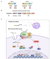
Schematic model for the roles in rMPT against MTB infection. (A) Domain screening of interacting site between MPT63 or MPT64 with TBK1, p47phox and HK2. (B) Construction of rMPT combined TBK1, p47phox, and HK2 interacting domains in MPT63 and MPT64. (C) Regulatory pathway of rMPT in macrophages.

Similar articles

Cited by

References

    1. Lechartier B., Rybniker J., Zumla A., Cole S.T. Tuberculosis drug discovery in the post-post-genomic era. EMBO Mol. Med. 2014;6:158–168. doi: 10.1002/emmm.201201772. - DOI - PMC - PubMed
    1. Bhatt K., Salgame P. Host innate immune response to Mycobacterium tuberculosis. J. Clin. Immunol. 2007;27:347–362. doi: 10.1007/s10875-007-9084-0. - DOI - PubMed
    1. Queval C.J., Brosch R., Simeone R. The Macrophage: A Disputed Fortress in the Battle against Mycobacterium tuberculosis. Front. Microbiol. 2017;8:2284. doi: 10.3389/fmicb.2017.02284. - DOI - PMC - PubMed
    1. Augenstreich J., Briken V. Host Cell Targets of Released Lipid and Secreted Protein Effectors of Mycobacterium tuberculosis. Front. Cell. Infect. Microbiol. 2020;10:595029. doi: 10.3389/fcimb.2020.595029. - DOI - PMC - PubMed
    1. Coombes B.K., Valdez Y., Finlay B.B. Evasive maneuvers by secreted bacterial proteins to avoid innate immune responses. Curr. Biol. 2004;14:R856–R867. doi: 10.1016/j.cub.2004.09.043. - DOI - PubMed