Review
. 2021 May 13;22(10):5154.
doi: 10.3390/ijms22105154.

Principles and Practical Considerations for the Analysis of Disease-Associated Alternative Splicing Events Using the Gateway Cloning-Based Minigene Vectors pDESTsplice and pSpliceExpress

Affiliations
Review

Principles and Practical Considerations for the Analysis of Disease-Associated Alternative Splicing Events Using the Gateway Cloning-Based Minigene Vectors pDESTsplice and pSpliceExpress

Elena Putscher et al. Int J Mol Sci. .

Abstract

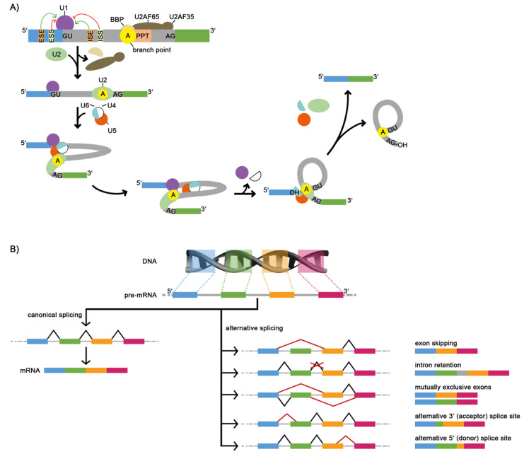
Splicing is an important RNA processing step. Genetic variations can alter the splicing process and thereby contribute to the development of various diseases. Alterations of the splicing pattern can be examined by gene expression analyses, by computational tools for predicting the effects of genetic variants on splicing, and by splicing reporter minigene assays for studying alternative splicing events under defined conditions. The minigene assay is based on transient transfection of cells with a vector containing a genomic region of interest cloned between two constitutive exons. Cloning can be accomplished by the use of restriction enzymes or by site-specific recombination using Gateway cloning. The vectors pDESTsplice and pSpliceExpress represent two minigene systems based on Gateway cloning, which are available through the Addgene plasmid repository. In this review, we describe the features of these two splicing reporter minigene systems. Moreover, we provide an overview of studies in which determinants of alternative splicing were investigated by using pDESTsplice or pSpliceExpress. The studies were reviewed with regard to the investigated splicing regulatory events and the experimental strategy to construct and perform a splicing reporter minigene assay. We further elaborate on how analyses on the regulation of RNA splicing offer promising prospects for gaining important insights into disease mechanisms.

Keywords: RNA processing; alternative splicing; gateway cloning; pDESTsplice; pSpliceExpress; splicing regulation; splicing reporter minigene assay.


Conflict of interest statement

The authors declare no conflict of interest in relation to this article.

Figures

Figure 1
Figure 1
The splicing process and common types of alternative splicing events (ASE). (A) Simplified scheme of pre-mRNA splicing with two exons (blue and green boxes) and one intron (gray box). The cis-regulatory elements, namely, 5′ and 3′ splice sites, which are evolutionarily highly conserved (usually GU and AG, respectively), branch point (yellow circle), polypyrimidine tract (light pink box), and splicing enhancers and silencers (ESE, ESS, ISE, ISS, light orange and light-yellow boxes) assist the spliceosome in recognizing the 5′- and 3′-ends of the intron. Positive regulation is indicated by green arrows, while negative regulation is shown in red. The formation of the spliceosome complex leads to conformational changes of the pre-mRNA. In the first step, the U1 snRNP binds to the GU sequence at the 5′ splice site. At the same time, the branch point is bound by the branch point-binding protein (BBP) and the polypyrimidine tract is bound by U2AF. In the next step, the BBP is replaced from the branch point by the U2 snRNP. The interaction of the branch point with U2 leads to the recruitment of the U4/U5/U6 snRNP complex and thereby to the formation of the pre-catalytic spliceosome. The following change of the spliceosome conformation leads to the release of U1 and U4. Then, the interaction of U6 with U2 results in a transesterification, where the guanosine of the 5′ splice site is bound to the adenosine in the branch point. In a second transesterification step, the exons are joined together. The spliced-out intron (lariat structure) is degraded, and the U2, U5, and U6 snRNPs are released to catalyze the following splicing process. (B) The canonical (left) and alternative (right) splicing paths with corresponding alternative splicing events that can be distinguished. The blue-, green-, orange-, and pink-colored boxes represent 4 different exons in the 5′ to 3′ direction, while the gray lines in between represent introns. The constitutive path of intron removal (black lines) and alternative paths (red lines) are indicated. ASE: alternative splicing events, BBP: branch point-binding protein, ESE: exonic splicing enhancer, ESS: exonic splicing silencer, ISE: intronic splicing enhancer, ISS: intronic splicing silencer, PPT: polypyrimidine tract, snRNP: small nuclear ribonucleoprotein, U2AF: U2 auxiliary factor.
Figure 2
Figure 2
Gateway cloning with pSpliceExpress and pDESTsplice. (A) The BP reaction is mediated by Int and IHF and leads to the cloning of the GFI (green box) into pSpliceExpress. Initially, the GFI (green box), which contains one or more alternatively spliced exons together with flanking intronic sequences, is surrounded by 25 bps long attB1 and attB2 sites (dark blue boxes). Important sequences of the pSpliceExpress vector are those of the ccdB gene (red box), the chloramphenicol resistance gene (white box), the attP1 and attP2 sites (orange boxes), the AmpR gene (light pink box), and the rat insulin 2 exons 2 and 3 (burgundy boxes). The resulting Expression clone contains the sequences of the attL1 and attL2 sites (orange and dark blue circles) and the GFI. The ccdB gene and the chloramphenicol resistance gene segments flanked by attR1 and attR2 sites (dark blue and orange circles) form the by-product of the BP reaction. (B) The LR reaction is mediated by Int, IHF, and Xis and leads to the cloning of the GFI into the pDESTsplice vector. Important sequence parts of the Entry clone are the GFI flanked by the attL1 and attL2 sites and an antibiotic resistance gene (e.g., for kanamycin resistance, purple box). The pDESTsplice vector principally contains the same sequence elements as the pSpliceExpress vector, except that pDESTsplice contains the attR sites instead of the attP sites. The resulting Expression clone contains the GFI flanked by the attB sites. The by-product is a vector containing the ccdB gene sequence and the chloramphenicol resistance gene sequence flanked by the attP sites. The sequences of attB1/B2, attP1/P2, attL1/L2, and attR1/R2 represent the forward strand only and are displayed in the same colors as in the schemes above. AmpR: ampicillin resistance, att: attachment, CmR: chloramphenicol resistance, GFI: genomic fragment of interest, IHF: integration host factor proteins, Int: integrase, Xis: excisionase.
Figure 3
Figure 3
Typical workflow for assessing the impact of a genetic variant on splicing by minigene assay. Example Expression clones are shown, where two variants of a GFI are cloned into pDESTsplice. The variants of the Expression clone contain GFIs, which here represent two allelic variants of a SNP, one of them originating from a nucleotide substitution of G to C (WT: G, green box and Mut: C, green/light blue box). Constitutive splicing is expected for WT and alternative splicing for Mut. E. coli are transformed with a mixture containing the Expression clone, the by-product, pDESTsplice, and the Entry clone. For the components of the different vectors, the same color pattern as in Figure 2 was used. Bacteria containing WT and Mut are efficiently selected due to the ampicillin resistance gene and the ccdB gene. The selection is illustrated by green ticks (bacteria survive) and red crosses (bacteria cannot survive). The plasmid DNA is amplified and isolated. The verification of WT and Mut can be performed by restriction enzyme digestion and sequencing of extracted DNA bands. WT and Mut Expression clones are used to transfect cells. The transfected cells are incubated usually for 24 to 48 h before the RNA is isolated. The RNA is used for RT-PCR. The PCR products of WT and Mut are confirmed by gel electrophoresis and by sequencing. In the illustrated example, the Mut genotype leads to preferential skipping of the investigated exon. DL: DNA ladder, GFI: genomic fragment of interest, Mut: mutant, RT-PCR: reverse transcriptase polymerase chain reaction, WT: wild-type.

Similar articles

Cited by

References

    1. Shi Y. Mechanistic insights into precursor messenger RNA splicing by the spliceosome. Nat. Rev. Mol. Cell Biol. 2017;18:655–670. doi: 10.1038/nrm.2017.86. - DOI - PubMed
    1. Scotti M.M., Swanson M.S. RNA mis-splicing in disease. Nat. Rev. Genet. 2016;17:19–32. doi: 10.1038/nrg.2015.3. - DOI - PMC - PubMed
    1. Park E., Pan Z., Zhang Z., Lin L., Xing Y. The expanding landscape of alternative splicing variation in human populations. Am. J. Hum. Genet. 2018;102:11–26. doi: 10.1016/j.ajhg.2017.11.002. - DOI - PMC - PubMed
    1. Kishore S., Khanna A., Stamm S. Rapid generation of splicing reporters with pSpliceExpress. Gene. 2008;427:104–110. doi: 10.1016/j.gene.2008.09.021. - DOI - PMC - PubMed
    1. Piovesan A., Caracausi M., Antonaros F., Pelleri M.C., Vitale L. GeneBase 1.1: A tool to summarize data from NCBI gene datasets and its application to an update of human gene statistics. Database. 2016;2016 doi: 10.1093/database/baw153. - DOI - PMC - PubMed

Substances