Review
. 2021 May 13;10(5):1191.
doi: 10.3390/cells10051191.

Novel Roles of SH2 and SH3 Domains in Lipid Binding

Affiliations
Review

Novel Roles of SH2 and SH3 Domains in Lipid Binding

Szabolcs Sipeki et al. Cells. .

Abstract

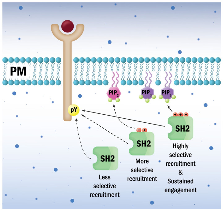
Signal transduction, the ability of cells to perceive information from the surroundings and alter behavior in response, is an essential property of life. Studies on tyrosine kinase action fundamentally changed our concept of cellular regulation. The induced assembly of subcellular hubs via the recognition of local protein or lipid modifications by modular protein interactions is now a central paradigm in signaling. Such molecular interactions are mediated by specific protein interaction domains. The first such domain identified was the SH2 domain, which was postulated to be a reader capable of finding and binding protein partners displaying phosphorylated tyrosine side chains. The SH3 domain was found to be involved in the formation of stable protein sub-complexes by constitutively attaching to proline-rich surfaces on its binding partners. The SH2 and SH3 domains have thus served as the prototypes for a diverse collection of interaction domains that recognize not only proteins but also lipids, nucleic acids, and small molecules. It has also been found that particular SH2 and SH3 domains themselves might also bind to and rely on lipids to modulate complex assembly. Some lipid-binding properties of SH2 and SH3 domains are reviewed here.

Keywords: SH2 domain; SH3 domain; Src; lipid binding; proline-rich sequences; protein tyrosine kinase.


Conflict of interest statement

The authors declare no conflict of interest.

Figures

Figure 1
Figure 1
Lipid-binding abilities of some SH2 domains may contribute to spatio-temporal coordination of tyrosine kinase signaling hub dynamics. Alternative cationic patches distinct from the phosphotyrosine-binding pockets serve as the primary binding site for membrane lipids in most SH2 domains attracted to lipids. Phosphatidylinositol-4,5-bisphosphate or phosphatidylinositol-3,4,5-trisphosphate are preferred over other phosphoinositides for binding by these elements. The shape and net charge of the respective alternative cationic patches of specific SH2 domains provide further lipid recognition bias and may guide their host proteins to distinct membrane regions.
Figure 2
Figure 2
Visualization of the interacting zones of certain SH2 domains. The cationic patches responsible for lipid binding (blue) in different SH2 domains do not overlap with the pockets employed for specific phosphotyrosine recognition (red). For the generation of the schematic representation of the SH2 domain, we used the RCSB PDB database (www.rcsb.org accessed on 3 May 2021).
Figure 3
Figure 3
Comparison of lipid binding SH3 domains. The prototype c-src SH3 domain employs key aromatic (red) and proline (yellow) residues for binding proline-rich motifs of their partner (green). The Caskin SH3 domain traded off the three key aromatic residues for basic (blue) and hydrophobic (white) ones. The hSH3 domain of ADAP features an N-terminal helix (bue) rich in basic amino acids. We used the RCSB PDB database (www.rcsb.org accessed on 3 May 2021) for the source of SH3 domain structure representations.
Figure 4
Figure 4
Some SH3 domains are involved in lipid recognition rather than protein binding. Typical SH3 domains accommodate two conserved pairs of aromatic/hydrophobic amino acids in two grooves for the attachment to two prolines in register (PXXP) on the surface of partner proteins. The helically extended SH3 (hSH3) domain identified in ADAP and PRAM-1 nests basic amino acids within an extra N-terminal helix. The composite surface created in the hSH3 domain displays cationic patches and can bind phosphatidylinositol-4,5-bisphosphate or phosphatidylinositol-3,4,5-trisphosphate but not proteins. The Caskin1 SH3 domain exchanged some proline-binding amino acids and lost the two PXXP motif-binding grooves. These changes resulted in a switch from protein to lipid binding ability. The Caskin1 SH3 domain preferentially binds to lysophosphatidic acid, a signaling-borne lipid found in positively charged membrane curvatures. Thus, hSH3 and Caskin1 SH3 domains might guide their host proteins along with their interacting partners to specific membrane sub-domains.

References

    1. Mayer B.J. The discovery of modular binding domains: Building blocks of cell signalling. Nat. Rev. Mol. Cell Biol. 2015;16:691–698. doi: 10.1038/nrm4068. - DOI - PubMed
    1. Sadowski I., Stone J.C., Pawson T. A noncatalytic domain conserved among cytoplasmic protein-tyrosine kinases modifies the kinase function and transforming activity of Fujinami sarcoma virus P130gag-fps. Mol. Cell. Biol. 1986;6:4396–4408. doi: 10.1128/MCB.6.12.4396. - DOI - PMC - PubMed
    1. Anderson D., Koch C.A., Grey L., Ellis C., Moran M.F., Pawson T. Binding of SH2 domains of phospholipase Cγ1, GAP, and Src to activated growth factor receptors. Science. 1990;250:979–982. doi: 10.1126/science.2173144. - DOI - PubMed
    1. Margolis B., Li N., Koch A., Mohammadi M., Hurwitz D.R., Zilberstein A., Ullrich A., Pawson T., Schlessinger J. The tyrosine phosphorylated carboxyterminus of the EGF receptor is a binding site for GAP and PLC-γ. EMBO J. 1990;9:4375–4380. doi: 10.1002/j.1460-2075.1990.tb07887.x. - DOI - PMC - PubMed
    1. Matsuda M., Mayer B.J., Hanafusa H. Identification of domains of the v-crk oncogene product sufficient for association with phosphotyrosine-containing proteins. Mol. Cell. Biol. 1991;11:1607–1613. doi: 10.1128/MCB.11.3.1607. - DOI - PMC - PubMed

Publication types

LinkOut - more resources