. 2021 May 13;10(5):771.
doi: 10.3390/antiox10050771.

Crosstalk between Peroxisomal Activities and Nrf2 Signaling in Porcine Embryos

Affiliations

Crosstalk between Peroxisomal Activities and Nrf2 Signaling in Porcine Embryos

Eui-Hyun Kim et al. Antioxidants (Basel). .

Abstract

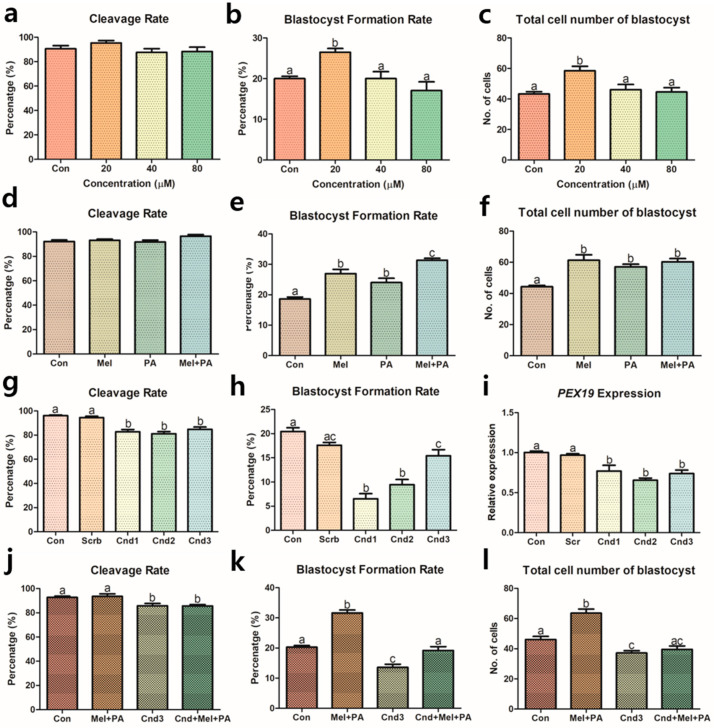
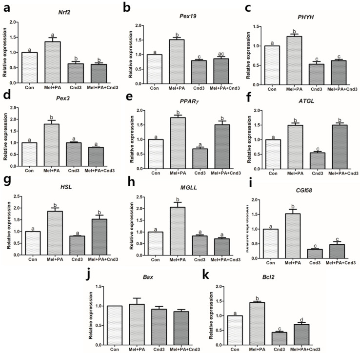
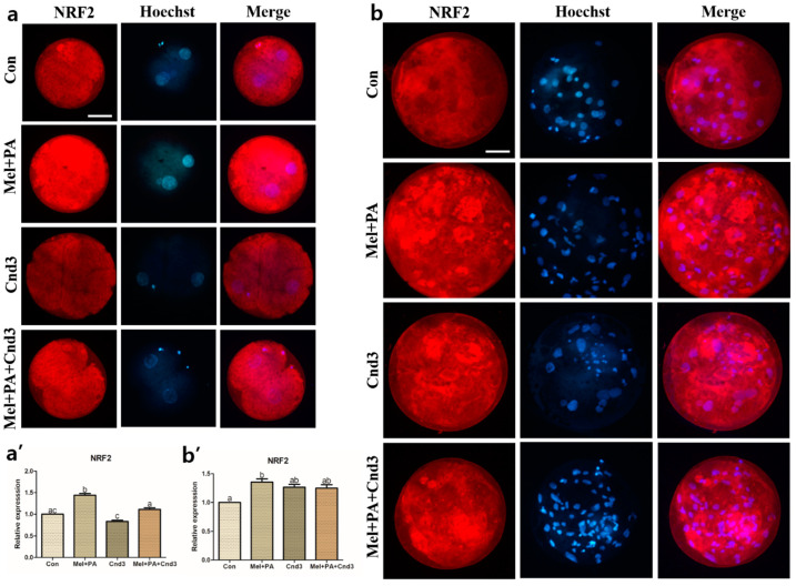
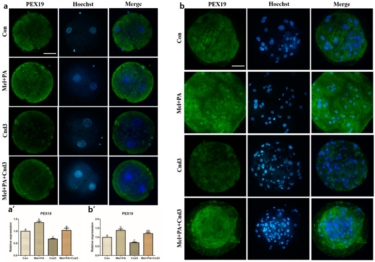
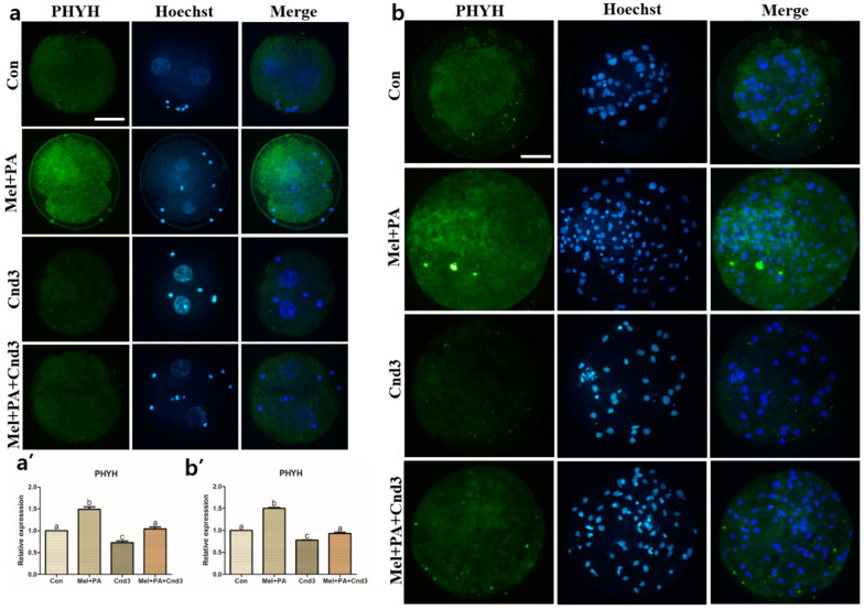
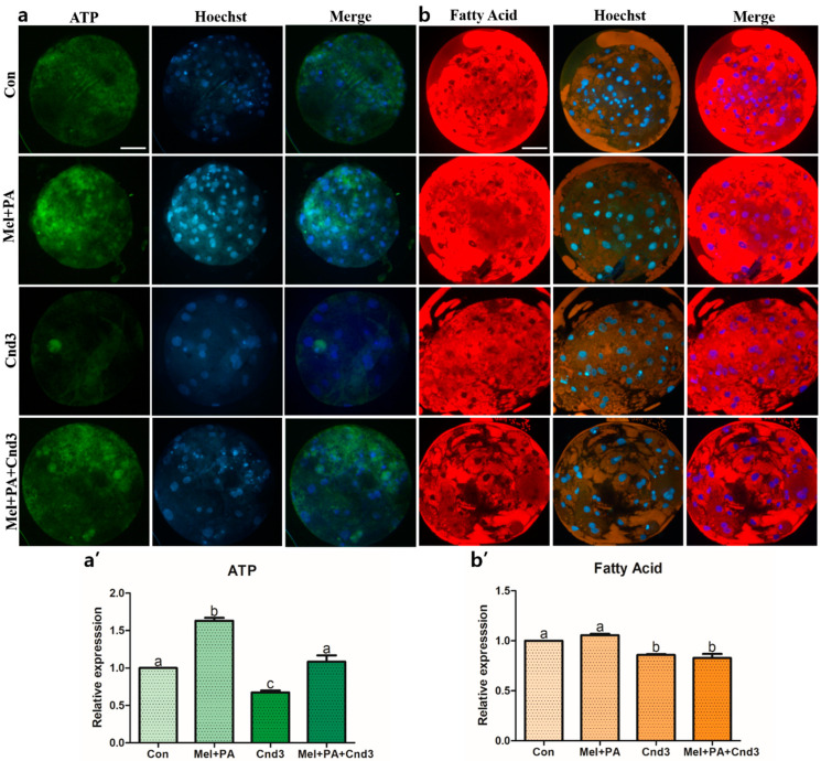
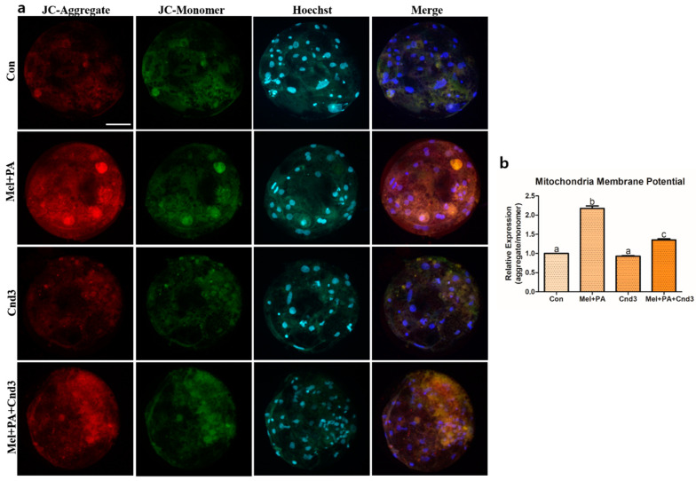
Melatonin and phytanic acid (PA) are known to be involved in lipid metabolism and β-oxidation, in which peroxisomal activities also significantly participate. In addition, other studies have reported that the nuclear factor-erythroid-derived 2-like 2 (Nrf2 or NFE2L2) signaling pathway mediates lipid metabolism and its subsequent cascades. As these mechanisms are partially involved in porcine oocytes or embryonic development, we hypothesized that the factors governing these mechanisms could be interconnected. Therefore, we aimed to investigate possible crosstalk between peroxisomal activities and Nrf2 signaling in porcine embryos following melatonin and PA treatment. Porcine embryos were cultured for seven days after parthenogenetic activation, and subsequently treated with melatonin and PA, or injected with Pex19-targeted siRNAs. Real-time PCR, immunocytochemistry, and BODIPY staining were used to evaluate peroxisomal activities, Nrf2 signaling, and subsequent lipid metabolism. We found that melatonin/PA treatment enhanced embryonic development, whereas injection with Pex19-targeted siRNAs had the opposite effect. Moreover, melatonin/PA treatment upregulated peroxisomal activities, Nrf2 signaling, lipid metabolism, and mitochondrial membrane potentials, whereas most of these mechanisms were downregulated by Pex19-targeted siRNAs. Therefore, we suggest that there is a connection between the action of melatonin and PA and the Nrf2 signaling pathway and peroxisomal activities, which positively influences porcine embryonic development.

Keywords: IVC; Nrf2 signaling; lipid metabolism; melatonin; phytanic acid; porcine embryos.


Conflict of interest statement

The authors declare that there is no conflict of interest.

Figures

Figure 1
Figure 1
Assessments of porcine embryonic development treated with melatonin and phytanic acid (PA), and injected with Pex19-targeted siRNAs. (ac) Optimization of PA during porcine IVC through rates of cleavage, blastocyst formation, and total cell numbers of blastocysts. (df) Assessment of co-treatment effect by melatonin and PA. (g,h) Selection of Pex19-targeted siRNAs through embryonic development and (i) verification of Pex19 knockdown by real-time PCR. (jl) Assessment of melatonin/PA treatment and siRNA on porcine embryos through rates of cleavage, blastocyst formation, and total cell numbers of blastocysts. At least five biological replications were performed and more than 170 embryos from each experimental group were used. Data are shown as the means ± SEM. Groups marked with different alphabetical letters are significantly different (p < 0.05). Con, control; mel, melatonin; PA, phytanic acid; Cnd, candidate.
Figure 2
Figure 2
Gene expression evaluations of melatonin/PA treatment and Cnd3-injection on porcine two-cell embryos. The gene expressions of (a) Nrf2, the main factor in the Nrf2 signaling pathway, (be) factors that are related to peroxisomal activities, (fi) lipid metabolism, and (j,k) apoptosis in the embryos were analyzed within the four groups. More than 200 embryos in each experimental group from six biological replications were used and real-time PCR was performed three times technically. Within the same mRNA, bars with different alphabetical letters are significantly different among the groups (p < 0.05). Con, control; mel, melatonin, PA, phytanic acid; Cnd3, siRNA candidate 3.
Figure 3
Figure 3
Gene expression evaluations of melatonin/PA treatment and Cnd3-injection on porcine blastocysts. The gene expressions of (a) Nrf2, the main factor in the Nrf2 signaling pathway, (be) factors that are related to peroxisomal activities, (fi) lipid metabolism, and (j,k) apoptosis in the embryos were analyzed within the four groups. More than 50 blastocysts in each experimental group from six biological replications were used and real-time PCR was performed three times technically. Within the same mRNA, bars with different alphabetical letters are significantly different among the groups (p < 0.05). Con, control; mel, melatonin, PA, phytanic acid; Cnd3, siRNA candidate 3.
Figure 4
Figure 4
Representative images of immunocytochemical analysis of NRF2 in porcine two-cell embryo blastocysts. Fluorescence microscopy was applied to obtain images of embryos. (a,b) Representative two-cell embryos in each group were stained with NRF2 rabbit-derived antibody and counterstained with Hoechst 33342. (a’,b’) Analysis of intensities from NRF2-stained two-cell embryos and blastocysts. At least 20 two-cell embryos and 15 blastocysts from five biological replications in each group were used for the staining and immunocytochemistry was performed three times technically. Data are shown as the means ± SEM. Bars with different alphabetical letters are significantly different (p < 0.05). Con, control; mel, melatonin, PA, phytanic acid; Cnd3, siRNA candidate 3. White bars in the images indicate 50 µm; 400× magnification.
Figure 5
Figure 5
Representative images of immunocytochemical analysis of PEX19 in porcine two-cell embryo blastocysts. Fluorescence microscopy was applied to obtain images of embryos. (a,b) Representative two-cell embryos in each group were stained with PEX19 rabbit-derived antibody and counterstained with Hoechst 33342. (a’,b’) Analysis of intensities from PEX19-stained two-cell embryos and blastocysts. At least 20 two-cell embryos and 15 blastocysts from five biological replications in each group were used for the staining and immunocytochemistry was performed three times technically. Data are shown as the means ± SEM. Bars with different alphabetical letters are significantly different (p < 0.05). Con, control; mel, melatonin, PA, phytanic acid; Cnd3, siRNA candidate 3. White bars in the images indicate 50 µm; 400× magnification.
Figure 6
Figure 6
Representative images of immunocytochemical analysis of PHYH in porcine two-cell embryo blastocysts. Fluorescence microscopy was applied to obtain images of embryos. (a,b) Representative two-cell embryos in each group were stained with PHYH rabbit-derived antibody and counterstained with Hoechst 33342. (a’,b’) Analysis of intensities from PHYH-stained two-cell embryos and blastocysts. At least 20 two-cell embryos and 15 blastocysts from five biological replications in each group were used for the staining and immunocytochemistry was performed three times technically. Data are shown as the means ± SEM. Bars with different alphabetical letters are significantly different (p < 0.05). Con, control; mel, melatonin, PA, phytanic acid; Cnd3, siRNA candidate 3. White bars in the images indicate 50 µm; 400× magnification.
Figure 7
Figure 7
Representative images of BODIPY staining analysis of ATP contents and fatty acids in blastocysts. Fluorescence microscopy was applied to obtain images of the blastocysts. (a,b) Representative blastocysts in each group were stained with BODIPY FL ATP and BODIPY 558/568 C12, respectively, then counter-stained with Hoechst 33342. (a’,b’) Analysis of intensities from BODIPY FL ATP- and BODIPY 558/568 C12-stained blastocysts. At least 15 blastocysts from four biological replications in each group were used for the staining and it was performed three times technically. Data are shown as the means ± SEM. Bars with different alphabetical letters are significantly different (p < 0.05). Con, control; mel, melatonin, PA, phytanic acid; Cnd3, siRNA candidate 3. White bars in the images indicate 50 µm; 400× magnification.
Figure 8
Figure 8
Representative images of JC-1 MMP staining in blastocysts. Fluorescence microscopy was applied to obtain images of the blastocysts. (a) Representative blastocysts in each group were stained with JC-1 aggregate and JC-1 monomer, then counter-stained with Hoechst 33342. (b) Analysis of intensities from JC-1 MMP-stained blastocysts. At least 15 blastocysts from four biological replications in each group were used for the staining and it was performed three times technically. Data are shown as the means ± SEM. Bars with different alphabetical letters are significantly different (p < 0.05). Con, control; mel, melatonin, PA, phytanic acid; Cnd3, siRNA candidate 3. White bars in the images indicate 50 µm; 400× magnification.

Similar articles

Cited by

References

    1. Aigner B., Renner S., Kessler B., Klymiuk N., Kurome M., Wünsch A., Wolf E. Transgenic pigs as models for translational biomedical research. J. Mol. Med. 2010;88:653–664. doi: 10.1007/s00109-010-0610-9. - DOI - PubMed
    1. Prather R.S., Hawley R.J., Carter D.B., Lai L., Greenstein J.L. Transgenic swine for biomedicine and agriculture. Theriogenology. 2003;59:115–123. doi: 10.1016/S0093-691X(02)01263-3. - DOI - PubMed
    1. Wu J., Vilarino M., Suzuki K., Okamura D., Bogliotti Y.S., Park I., Rowe J., McNabb B., Ross P.J., Belmonte J.C. CRISPR-Cas9 mediated one-step disabling of pancreatogenesis in pigs. Sci. Rep. 2017;7:10487. doi: 10.1038/s41598-017-08596-5. - DOI - PMC - PubMed
    1. Brussow K.P., Torner H., Kanitz W., Ratky J. In vitro technologies related to pig embryo transfer. Reprod. Nutr. Dev. 2000;40:469–480. doi: 10.1051/rnd:2000111. - DOI - PubMed
    1. Dang-Nguyen T.Q., Wells D., Haraguchi S., Men N.T., Nguyen H.T., Noguchi J., Kaneko H., Kikuchi K. Combined refinements to somatic cell nuclear transfer methods improve porcine embryo development. J. Reprod. Dev. 2020;66:281–286. doi: 10.1262/jrd.2019-156. - DOI - PMC - PubMed