. 2021 May 13;10(5):1194.
doi: 10.3390/cells10051194.

Metabolomic Analysis Evidences That Uterine Epithelial Cells Enhance Blastocyst Development in a Microfluidic Device

Affiliations

Metabolomic Analysis Evidences That Uterine Epithelial Cells Enhance Blastocyst Development in a Microfluidic Device

Vanessa Mancini et al. Cells. .

Abstract

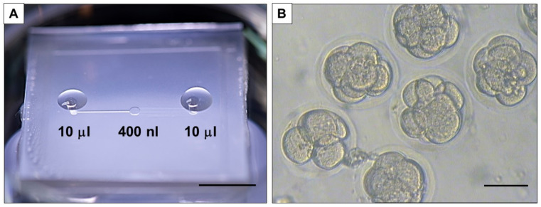
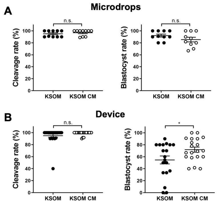
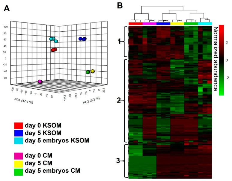
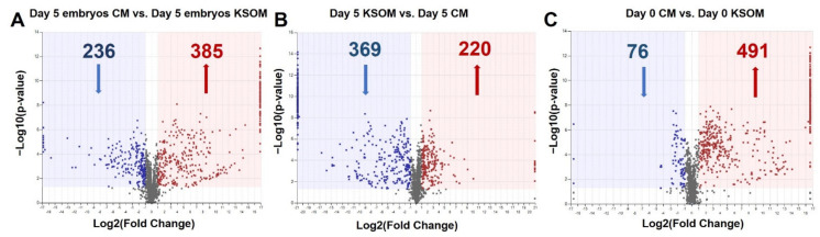
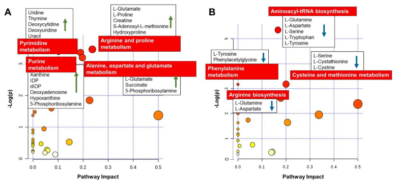
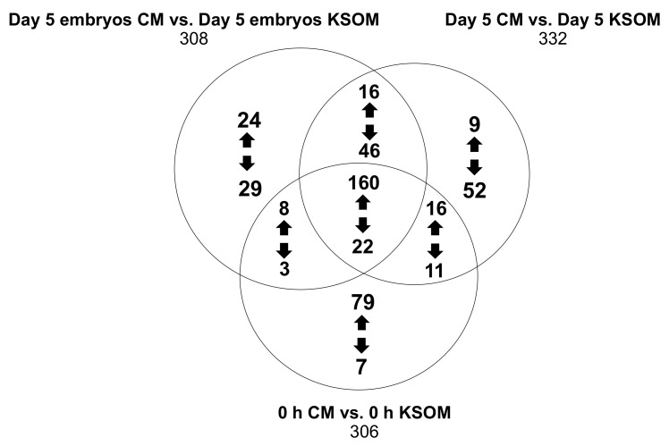
Here we report the use of a microfluidic system to assess the differential metabolomics of murine embryos cultured with endometrial cells-conditioned media (CM). Groups of 10, 1-cell murine B6C3F1 × B6D2F1 embryos were cultured in the microfluidic device. To produce CM, mouse uterine epithelial cells were cultured in potassium simplex optimized medium (KSOM) for 24 h. Media samples were collected from devices after 5 days of culture with KSOM (control) and CM, analyzed by reverse phase liquid chromatography and untargeted positive ion mode mass spectrometry analysis. Blastocyst rates were significantly higher (p < 0.05) in CM (71.8%) compared to control media (54.6%). We observed significant upregulation of 341 compounds and downregulation of 214 compounds in spent media from CM devices when compared to control. Out of these, 353 compounds were identified showing a significant increased abundance of metabolites involved in key metabolic pathways (e.g., arginine, proline and pyrimidine metabolism) in the CM group, suggesting a beneficial effect of CM on embryo development. The metabolomic study carried out in a microfluidic environment confirms our hypothesis on the potential of uterine epithelial cells to enhance blastocyst development. Further investigations are required to highlight specific pathways involved in embryo development and implantation.

Keywords: embryo culture; metabolomics; microfluidics.


Conflict of interest statement

The authors declare no conflict of interest.

Figures

Figure 1
Figure 1
Microfluidic device. (A) The device is placed in a 60 mm petri dish for loading medium and kept in the incubator for the duration of the culture (scale bar 1 cm). (B) The developing embryos visualized in the central chamber in brightfield (scale bar 100 µm).
Figure 2
Figure 2
Effects of CM on embryo development. Cleavage rates (left) and blastocyst rates (right) of embryos cultured in drops (A) or in microfluidic devices (B). Each dot represents an individual culture, in which 10 embryos are cultured. Data presented as mean ± SEM. Non significant (n.s.) differences and significant (*) comparison are shown (* p < 0.05).
Figure 3
Figure 3
The metabolite composition of the media used for embryo culture is considerably changed by uterine epithelial cells. LC-MS/MS global metabolomic profile analysis of blastocysts cultured in KSOM or CM in devices. (A) PCA plot for LC-MS/MS data of medium samples collected from devices in different experimental conditions (n = 3 for each group): spent KSOM (blue) or CM (yellow) from devices without embryos, spent KSOM (cyan) or CM (green) from devices with embryos and the relative controls. (B) Heatmap representing LC-MS/MS data. Compounds are presented as rows and sample replicates as columns and processed by using Euclidean distance and Ward clustering via Metaboanalyst 4.0 for Pareto scaled, log transformed, and averaged group data. Colours are displayed by normalized abundance, ranging from low (green) to high (red).
Figure 4
Figure 4
Volcano plot showing the distribution of metabolic compounds for the pairwise comparison. Pairwise comparison of (A) day 5 embryo culture CM vs. day 5 embryo culture KSOM, (B) Day 5 CM vs. Day 5 KSOM, and (C) Day 0 CM vs. Day 0 KSOM. Significance criteria: p ≤ 0.05, fold change ≥ |2|. Volcano plots combine the p-value measured by ANOVA, expressed as −log10(p-value), with the magnitude of the change in relative abundance, expressed as log2(fold change), between the groups considered in a particular pairwise comparison.
Figure 5
Figure 5
Metabolic pathways of significantly changed metabolites between day 5 embryo culture CM and day 5 embryo culture KSOM. (A) Increased metabolites uniquely produced by embryos when exposed to uterine epithelial cells. The graph presents a list of the matched overrepresented pathways for compounds significantly increased in day 5 embryo culture CM, compared to day 5 embryo culture KSOM, arranged by p-values on Y-axis, and pathway impact values on X-axis. The enriched pathways for the 208 increased metabolites identified in the CM group are represented by nodes of different size and color gradient corresponding to the significance of the pathway ranked by impact score (the larger the circle the higher the impact score) and p-value (yellow: higher p-values and red: lower p-values), respectively. The red labels highlight the main pathways. Five of these pathways had a p-value < 0.1 and 16 had pathway impact value higher than 0, which are the cut-off values for relevance. (B) Decreased metabolites uniquely produced by embryos when exposed to uterine epithelial cells. Only pathways with p-value < 0.1 were considered significant. Of these pathways, 10 had impact value higher than 0 and 10 had a p value < 0.2. The most significantly affected (p < 0.1) metabolic pathways are labelled and represented with larger red dots.
Figure 6
Figure 6
Venn diagram showing the distribution of the number of metabolites between the three pairwise comparisons.

Similar articles

Cited by

References

    1. Kupka M.S., Ferraretti A.P., De Mouzon J., Erb K., D’Hooghe T., Castilla J.A., Calhaz-Jorge C., De Geyter C., Goossens V., Strohmer H., et al. Assisted reproductive technology in Europe, 2010: Results generated from European registers by ESHRE. Hum. Reprod. 2014;29:2099–2113. doi: 10.1093/humrep/deu175. - DOI - PubMed
    1. Ascari I.J., Martins S.C., Camargo L.S.A., Mendez-Otero R. Development of bovine embryos in vitro in coculture with murine mesenchymal stem cells and embryonic fibroblasts. Mol. Biol. Rep. 2018;45:1827–1837. doi: 10.1007/s11033-018-4329-y. - DOI - PubMed
    1. Bavister B.D. Role of oviductal secretions in embryonic growth in vivo and in vitro. Theriogenology. 1988;29:143–154. doi: 10.1016/0093-691X(88)90037-4. - DOI
    1. Gardner D.K., Sakkas D. Mouse embryo cleavage, metabolism and viability: Role of medium composition. Hum. Reprod. 1993;8:288–295. doi: 10.1093/oxfordjournals.humrep.a138039. - DOI - PubMed
    1. Gardner D.K., Leese H.J. Concentrations of nutrients in mouse oviduct fluid and their effects on embryo development and metabolism in vitro. J. Reprod. Fertil. 1990;88:361–368. doi: 10.1530/jrf.0.0880361. - DOI - PubMed

Publication types

Substances

LinkOut - more resources