Review
. 2021 May 13;10(5):1196.
doi: 10.3390/cells10051196.

The Emerging Role of CT-Based Imaging in Adipose Tissue and Coronary Inflammation

Affiliations
Review

The Emerging Role of CT-Based Imaging in Adipose Tissue and Coronary Inflammation

Jeremy Yuvaraj et al. Cells. .

Abstract

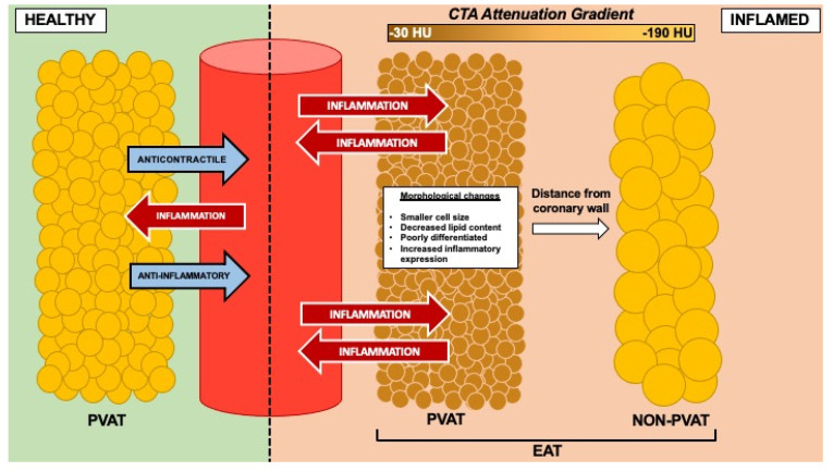
A large body of evidence arising from recent randomized clinical trials demonstrate the association of vascular inflammatory mediators with coronary artery disease (CAD). Vascular inflammation localized in the coronary arteries leads to an increased risk of CAD-related events, and produces unique biological alterations to local cardiac adipose tissue depots. Coronary computed tomography angiography (CTA) provides a means of mapping inflammatory changes to both epicardial adipose tissue (EAT) and pericoronary adipose tissue (PCAT) as independent markers of coronary risk. Radiodensity or attenuation of PCAT on coronary CTA, notably, provides indirect quantification of coronary inflammation and is emerging as a promising non-invasive imaging implement. An increasing number of observational studies have shown robust associations between PCAT attenuation and major coronary events, including acute coronary syndrome, and 'vulnerable' atherosclerotic plaque phenotypes that are associated with an increased risk of the said events. This review outlines the biological characteristics of both EAT and PCAT and provides an overview of the current literature on PCAT attenuation as a surrogate marker of coronary inflammation.

Keywords: adipose tissue; atherosclerosis; computed tomography coronary angiography; coronary artery disease; coronary inflammation; epicardial adipose tissue; pericoronary adipose tissue.


Conflict of interest statement

The authors declare no conflict of interest.

Figures

Figure 1
Figure 1
Schematic representation of bidirectional signalling between the vasculature and adipose tissue, particularly perivascular (or pericoronary) fractions of epicardial adipose tissue (EAT). Under ‘healthy’ conditions, perivascular adipose tissue (PVAT) typically exhibits anticontractile and anti-inflammatory properties in response to markers of oxidative stress and inflammation from the vasculature. Under ‘inflamed’ conditions, PVAT undergoes unique phenotypic changes due to vascular inflammation compared to non-perivascular adipose tissue located further from the vessel wall. These changes are detectable as increased attenuation on coronary computed tomography angiography (CTA).
Figure 2
Figure 2
Epicardial adipose tissue (EAT) shown in purple on axial view of coronary computed tomography angiography (CTA). EAT in patient without coronary artery disease (CAD) shown in left panel (A), and EAT in patient with CAD shown in right panel (B).
Figure 3
Figure 3
Pericoronary adipose tissue (PCAT) shown in cross-sectional (A,B) and longitudinal (C,D) views of the right coronary artery (RCA) on coronary computed tomography angiography (CTA). PCAT in RCA without plaque represented in the left panels (A,C), and PCAT in RCA with calcified and non-calcified plaque represented in the right panels (B,D). Colour map describes spectrum of adipose tissue attenuation values in Hounsfield units (HU), ranging from −190 HU (yellow) to −30 HU (red), with higher attenuation values indicating inflammatory changes.

References

    1. Ross R. Atherosclerosis is an inflammatory disease. Am. Heart J. 1999;138:S419–S420. doi: 10.1016/S0002-8703(99)70266-8. - DOI - PubMed
    1. Libby P. Inflammation in atherosclerosis. Arterioscler. Thromb. Vasc. Biol. 2012;32:2045–2051. doi: 10.1161/ATVBAHA.108.179705. - DOI - PMC - PubMed
    1. Moore K.J., Koplev S., Fisher E.A., Tabas I., Bjorkegren J.L.M., Doran A.C., Kovacic J.C. Macrophage Trafficking, Inflammatory Resolution, and Genomics in Atherosclerosis: JACC Macrophage in CVD Series (Part 2) J. Am. Coll. Cardiol. 2018;72:2181–2197. doi: 10.1016/j.jacc.2018.08.2147. - DOI - PMC - PubMed
    1. Clinton S.K., Underwood R., Hayes L., Sherman M.L., Kufe D.W., Libby P. Macrophage colony-stimulating factor gene expression in vascular cells and in experimental and human atherosclerosis. Am. J. Pathol. 1992;140:301–316. - PMC - PubMed
    1. Hedrick C.C. Lymphocytes in atherosclerosis. Arterioscler. Thromb. Vasc. Biol. 2015;35:253–257. doi: 10.1161/ATVBAHA.114.305144. - DOI - PMC - PubMed

MeSH terms