

Mitochondrial Bioenergetics and Turnover during Chronic Muscle Disuse
- PMID: 34068411
- PMCID: PMC8153634
- DOI: 10.3390/ijms22105179
Mitochondrial Bioenergetics and Turnover during Chronic Muscle Disuse
Abstract
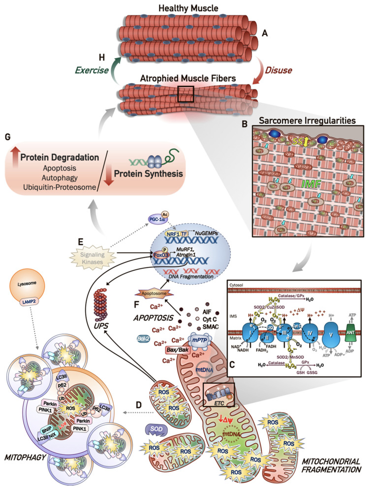
Periods of muscle disuse promote marked mitochondrial alterations that contribute to the impaired metabolic health and degree of atrophy in the muscle. Thus, understanding the molecular underpinnings of muscle mitochondrial decline with prolonged inactivity is of considerable interest. There are translational applications to patients subjected to limb immobilization following injury, illness-induced bed rest, neuropathies, and even microgravity. Studies in these patients, as well as on various pre-clinical rodent models have elucidated the pathways involved in mitochondrial quality control, such as mitochondrial biogenesis, mitophagy, fission and fusion, and the corresponding mitochondrial derangements that underlie the muscle atrophy that ensues from inactivity. Defective organelles display altered respiratory function concurrent with increased accumulation of reactive oxygen species, which exacerbate myofiber atrophy via degradative pathways. The preservation of muscle quality and function is critical for maintaining mobility throughout the lifespan, and for the prevention of inactivity-related diseases. Exercise training is effective in preserving muscle mass by promoting favourable mitochondrial adaptations that offset the mitochondrial dysfunction, which contributes to the declines in muscle and whole-body metabolic health. This highlights the need for further investigation of the mechanisms in which mitochondria contribute to disuse-induced atrophy, as well as the specific molecular targets that can be exploited therapeutically.
Keywords: apoptosis; autophagy; mitochondrial biogenesis; mitochondrial quality control; mitophagy; muscle disuse; reactive oxygen species; skeletal muscle atrophy.
Conflict of interest statement
The authors declare no conflict of interest.
Figures
Similar articles
-
Mitochondrial dysfunction induces muscle atrophy during prolonged inactivity: A review of the causes and effects.Arch Biochem Biophys. 2019 Feb 15;662:49-60. doi: 10.1016/j.abb.2018.11.005. Epub 2018 Nov 16. Arch Biochem Biophys. 2019. PMID: 30452895 Free PMC article. Review.
-
Regulation of mitochondrial quality following repeated bouts of hindlimb unloading.Appl Physiol Nutr Metab. 2020 Mar;45(3):264-274. doi: 10.1139/apnm-2019-0218. Epub 2019 Jul 24. Appl Physiol Nutr Metab. 2020. PMID: 31340136 Free PMC article.
-
p53 regulates skeletal muscle mitophagy and mitochondrial quality control following denervation-induced muscle disuse.J Biol Chem. 2022 Feb;298(2):101540. doi: 10.1016/j.jbc.2021.101540. Epub 2021 Dec 25. J Biol Chem. 2022. PMID: 34958797 Free PMC article.
-
Beneficial effects of exercise on age-related mitochondrial dysfunction and oxidative stress in skeletal muscle.J Physiol. 2016 Sep 15;594(18):5105-23. doi: 10.1113/JP270659. Epub 2015 Nov 21. J Physiol. 2016. PMID: 26503074 Free PMC article. Review.
-
Mitochondrial signaling contributes to disuse muscle atrophy.Am J Physiol Endocrinol Metab. 2012 Jul 1;303(1):E31-9. doi: 10.1152/ajpendo.00609.2011. Epub 2012 Mar 6. Am J Physiol Endocrinol Metab. 2012. PMID: 22395111 Free PMC article. Review.
Cited by
-
Guilu Erxian Jiao enhances protein synthesis, glucose homeostasis, mitochondrial biogenesis and slow-twitch fibers in the skeletal muscle.J Food Drug Anal. 2023 Mar 15;31(1):116-136. doi: 10.38212/2224-6614.3435. J Food Drug Anal. 2023. PMID: 37224559 Free PMC article.
-
PROKR1-CREB-NR4A2 axis for oxidative muscle fiber specification and improvement of metabolic function.Proc Natl Acad Sci U S A. 2024 Jan 23;121(4):e2308960121. doi: 10.1073/pnas.2308960121. Epub 2024 Jan 17. Proc Natl Acad Sci U S A. 2024. PMID: 38232288 Free PMC article.
-
Beeting atrophy: dietary nitrate to protect the powerhouse of the cell?J Physiol. 2025 Jul;603(13):3861-3862. doi: 10.1113/JP285115. Epub 2023 Jul 31. J Physiol. 2025. PMID: 37519113 Free PMC article. No abstract available.
-
Protective Effects of the Chalcone-Based Derivative AN07 on Inflammation-Associated Myotube Atrophy Induced by Lipopolysaccharide.Int J Mol Sci. 2022 Oct 26;23(21):12929. doi: 10.3390/ijms232112929. Int J Mol Sci. 2022. PMID: 36361718 Free PMC article.
-
Alcohol Alters Skeletal Muscle Bioenergetic Function: A Scoping Review.Int J Mol Sci. 2024 Nov 15;25(22):12280. doi: 10.3390/ijms252212280. Int J Mol Sci. 2024. PMID: 39596345 Free PMC article.
References
Publication types
MeSH terms
Grants and funding
LinkOut - more resources
Full Text Sources
