Case Reports
. 2021 May 13;22(10):5180.
doi: 10.3390/ijms22105180.

CACNA1A Mutations Causing Early Onset Ataxia: Profiling Clinical, Dysmorphic and Structural-Functional Findings

Affiliations
Case Reports

CACNA1A Mutations Causing Early Onset Ataxia: Profiling Clinical, Dysmorphic and Structural-Functional Findings

Antonio F Martínez-Monseny et al. Int J Mol Sci. .

Abstract

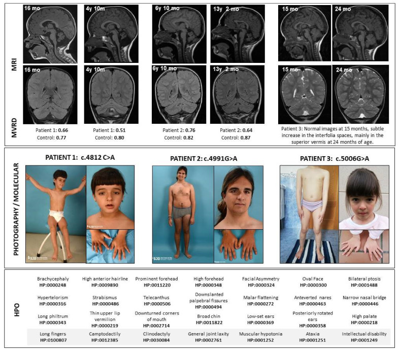
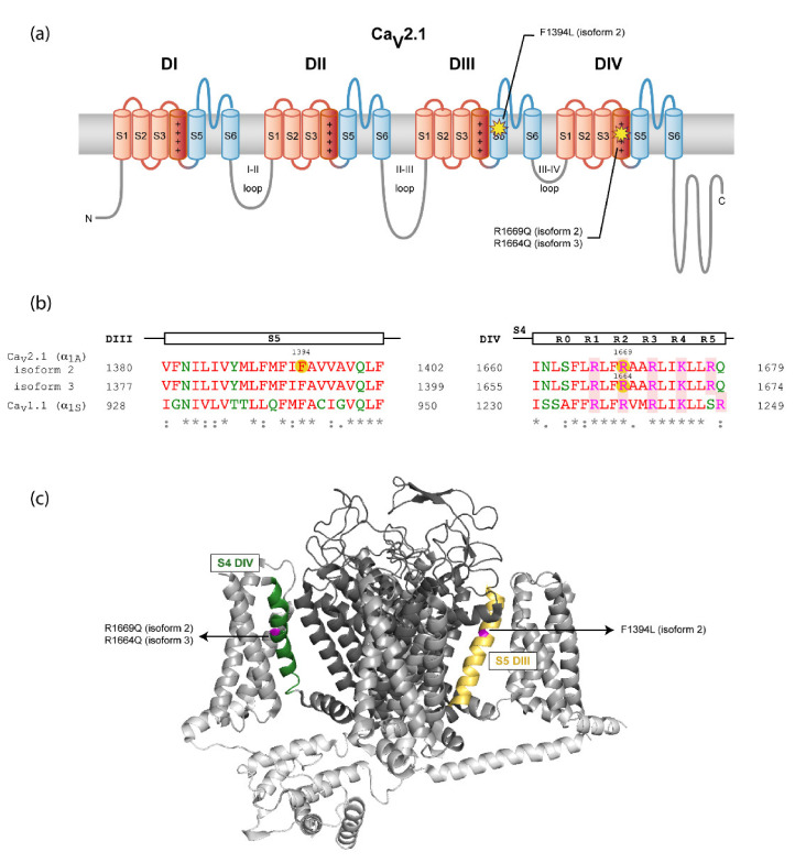
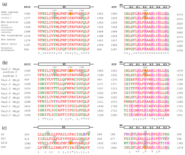
The CACNA1A gene encodes the pore-forming α1A subunit of the voltage-gated CaV2.1 Ca2+ channel, essential in neurotransmission, especially in Purkinje cells. Mutations in CACNA1A result in great clinical heterogeneity with progressive symptoms, paroxysmal events or both. During infancy, clinical and neuroimaging findings may be unspecific, and no dysmorphic features have been reported. We present the clinical, radiological and evolutionary features of three patients with congenital ataxia, one of them carrying a new variant. We report the structural localization of variants and their expected functional consequences. There was an improvement in cerebellar syndrome over time despite a cerebellar atrophy progression, inconsistent response to acetazolamide and positive response to methylphenidate. The patients shared distinctive facial gestalt: oval face, prominent forehead, hypertelorism, downslanting palpebral fissures and narrow nasal bridge. The two α1A affected residues are fully conserved throughout evolution and among the whole human CaV channel family. They contribute to the channel pore and the voltage sensor segment. According to structural data analysis and available functional characterization, they are expected to exert gain- (F1394L) and loss-of-function (R1664Q/R1669Q) effect, respectively. Among the CACNA1A-related phenotypes, our results suggest that non-progressive congenital ataxia is associated with developmental delay and dysmorphic features, constituting a recognizable syndromic neurodevelopmental disorder.

Keywords: CACNA1A gene; CaV2.1 (P/Q-type) voltage-dependent calcium channel; ataxia; cerebellar atrophy; dysmorphic traits; early-onset cerebellar ataxia.


Conflict of interest statement

The authors declare no conflict of interest.

Figures

Figure 1
Figure 1
Clinical and radiological features of patients. Above, the magnetic resonance sagittal and coronal images show a progression in the cerebellar atrophy in Patients 1 and 2, despite clinical stabilization. Immediately below the images, the midsagittal vermis relative diameter (MVRD) has been calculated in the sagittal sequences for patients 1 and 2. MVRDs are detailed and compared to controls’ values. In the middle, pictures from the patients are shown. In the bottom, Human Phenotype Ontology (HPO) codes are included. y: years; mo: months; MVRD: midsagittal vermis relative diameter.
Figure 2
Figure 2
Location of the mutations in CaV2.1 α1A channel subunit. (a) Location of the variant residues of α1A channel subunit isoforms 2 and 3 in the secondary structure of the protein according to the cryo-electron microscopy (cryo-EM) structure of the rabbit CaV1.1 complex, containing the pore-forming α1S and several regulatory subunits [12] (see also panel B). N- and C-termini and the intracellular loops are shown in gray, voltage-sensor modules (S1–S4) from the four domains are in red—with the S4 α-helixes colored in dark red—and Ca2+-selective pore modules (S5-P loop-S6) are in blue. (b) Sequence alignment of the regions affected by variants between the human α1A subunit isoforms 2 and 3 and rabbit CaV1.1 α1S subunit. The α1A subunit segment of each alignment is indicated at the top. Mutations are highlighted with yellow circles on the human CaV2.1 (hCaV2.1) sequences. The gating charged residues of S4 segments (labeled R0-R5) are shaded in red. Amino acids are colored depending on their physicochemical properties: small and hydrophobic are in red, acidic in blue, basic in magenta and G and amino acids containing hydroxyl, sulfhydryl or amine groups in green. Below, a consensus code indicates fully conserved residues (*), conservation between residues with strongly similar properties (:) or with weakly similar properties (.). The Uniport IDs of the sequences aligned are for hCaV2 (α1A): O00555-2 (isoform 2) and O00555-3 (isoform 3) and for rabbit CaV1.1 (α1S): P07293. The sequence alignments were made using Multiple Sequence Aligment Clustal Omega. (c) Three-dimensional location of the amino acid variants on a CaV channel model. The structure of the α1S subunit of the CaV1.1 channel (PDB 5GJV) [12] was used as a model considering their high level of homology (panel B). N- and C-termini and the intracellular loops are shown in light gray, voltage-sensor modules (S1–S4) are in gray and Ca2+-selective pore modules (S5-P loop-S6) are in dark gray. The two regions where mutations are located are highlighted in yellow for S5 helix of DIII and green for S4 helix of DIV. The residues of CaV1.1 (α1S) equivalent to those mutated in CaV2.1 (α1A) and identified in the patients, according to the sequence alignment on panel B, have been highlighted in magenta.
Figure 3
Figure 3
Sequence alignment of the α1A subunit regions affected by the amino acid variants. Sequence alignment of the α1A subunit regions affected by mutations, including α1A isoforms 2 and 3, between different species (a), between the human CaV channel family (b) and between the four domains of α1A subunit isoform 2 (c). The α1A region of each alignment is indicated at the top. CaV2.1 mutations are highlighted with yellow circles on the α1A subunit isoform 2 or 3. Other CaV2.1 residues affected by mutations mentioned in the text are indicated in gray circles. The gating charged amino acids of S4 segments (labeled R0-R5) and shaded in red. Residues are colored given their physicochemical properties: small and hydrophobic are in red, acidic in blue, basic in magenta and G and residues containing hydroxyl, sulfhydryl or amine groups in green. The consensus code below indicates fully conserved residues (*), conservative (:) or semi-conservative amino acid substitutions (.). The Uniport IDs of the aligned orthologous CaV2.1 sequences are: Mus musculus: P97445; Rattus norvegicus: P54282; Oryctolagus cuniculus: P27884; Pan troglodytes: A0A2I3T217; Bos taurus: F1N1E0; Danio rerio: E9QJF6; Drosophila melanogaster: P91645. The IDs of the human CaV channel family aligned are the following hCav1.1: Q13698; hCaV1.2: Q13936; hCaV1.3: Q01668; hCaV1.4: O60840; hCaV2.2: Q00975; hCaV2.3: Q15878; hCaV3.1: O43497; hCaV3.2: O95180; hCaV3.3: Q9P0X4. The sequence alignments were made using Multiple Sequence Aligment Clustal Omega. Here, residue numbers are indicated according to their position in α1A subunit isoform 2 and 3; however, some of them differ from the nomenclature of the mutations found in the literature: R1350Q/R1349Q corresponds to R1352Q in isoform 2, Y1385C to Y1387C in isoform 2 and Y1384C in isoform 3, V1396M is V1399M in isoform 2 and V1695I is V1700I in isoform 2.
Figure 4
Figure 4
Internal CaV1.1 channel amino acid interactions affected by mutations F942L and V947M. Structure of the rabbit CaV1.1 channel showing the side-chains of F942 and V947 and Yasara-identifed interacting residues from side (a) and top (b) views. Residues L938 and M941 are hidden in panel B for visualization purposes. DII S1-S2 loop and DIII S5, P-loop and S6 are colored in red, blue, green and yellow, respectively.

References

    1. Pietrobon D. CaV2.1 channelopathies. Pflügers Arch. Eur. J. Physiol. 2010;460:375–393. doi: 10.1007/s00424-010-0802-8. - DOI - PubMed
    1. Catterall W.A. Ion channel voltage sensors: Structure, function, and pathophysiology. Neuron. 2010;67:915–928. doi: 10.1016/j.neuron.2010.08.021. - DOI - PMC - PubMed
    1. Dolphin A.C. Voltage-gated calcium channels and their auxiliary subunits: Physiology and pathophysiology and pharmacology. J. Physiol. 2016;594:5369–5390. doi: 10.1113/JP272262. - DOI - PMC - PubMed
    1. Blumkin L., Michelson M., Leshinsky-Silver E., Kivity S., Lev D., Lerman-Sagie T. congenital ataxia, mental retardation, and dyskinesia associated with a novel CACNA1A mutation. J. Child Neurol. 2010;25:892–897. doi: 10.1177/0883073809351316. - DOI - PubMed
    1. Calandriello L., Veneziano L., Francia A., Sabbadini G., Colonnese C., Mantuano E., Jodice C., Trettel F., Viviani P., Manfredi M., et al. Acetazolamide-responsive episodic ataxia in an Italian family refines gene mapping on chromosome 19p13. Brain. 1997;120:805–812. doi: 10.1093/brain/120.5.805. - DOI - PubMed

Publication types