. 2021 May 10;13(5):871.
doi: 10.3390/v13050871.

Avian Influenza H7N9 Virus Adaptation to Human Hosts

Affiliations

Avian Influenza H7N9 Virus Adaptation to Human Hosts

Swan Tan et al. Viruses. .

Abstract

Avian influenza virus A (H7N9), after circulating in avian hosts for decades, was identified as a human pathogen in 2013. Herein, amino acid substitutions possibly essential for human adaptation were identified by comparing the 4706 aligned overlapping nonamer position sequences (1-9, 2-10, etc.) of the reported 2014 and 2017 avian and human H7N9 datasets. The initial set of virus sequences (as of year 2014) exhibited a total of 109 avian-to-human (A2H) signature amino acid substitutions. Each represented the most prevalent substitution at a given avian virus nonamer position that was selectively adapted as the corresponding index (most prevalent sequence) of the human viruses. The majority of these avian substitutions were long-standing in the evolution of H7N9, and only 17 were first detected in 2013 as possibly essential for the initial human adaptation. Strikingly, continued evolution of the avian H7N9 virus has resulted in avian and human protein sequences that are almost identical. This rapid and continued adaptation of the avian H7N9 virus to the human host, with near identity of the avian and human viruses, is associated with increased human infection and a predicted greater risk of human-to-human transmission.

Keywords: H7N9; adaptation; avian viruses; diversity; host; human viruses; influenza virus; motifs; surveillance; zoonosis.


Conflict of interest statement

We declare that we have no competing interests in this research.

Figures

Figure 1
Figure 1
Protein sequence diversity of avian and human influenza A (H7N9) viruses. Shannon’s entropy was used as a general measure of protein sequence diversity for each aligned nonamer (nine amino acids) position of the H7N9 avian (upper) and human (lower) virus proteomes. The entropy values indicate the level of variability at the corresponding nonamer positions, with a zero representing completely conserved sites and high entropy values of about 3 or higher marking highly variable sites.
Figure 2
Figure 2
Avian-to-human (A2H) substitution identified in the proteins of influenza A (H7N9) viruses. The amino acid positions of the A2H substitutions are indicated in the circles, and those underlined are the 50 that remained unchanged in the recorded human H7N9 population. The circles in green shade are substitutions that occurred in the evolutionary path of A (H7N9) viruses [16]; while those in yellow were first detected in 2013. The protein numeration is based on protein sequence alignment. Abbreviations: RdRp CS, RdRp catalytic subunit; HA, hemagglutinin; VS, virion surface; MB, membrane binding; RNPB, ribonucleoprotein binding; NLS, nuclear localization signal; SAMP (III), signal-anchor for type III membrane protein; IV, intravirion; THF, transmembrane helical fragments; RNABH, RNA-binding and homodimerization; CPSF4B, cleavage and polyadenylation specificity factor 4 binding; and NES, nuclear export signal.
Figure 3
Figure 3
Heat map depicting the distribution of the 109 identified avian-to-human (A2H) substitution sites (rows) of publicly reported, full-length, avian and human influenza A (H7N9) virus strains (columns). The identified A2H amino acid (a.a.) substitutions are sorted according to the influenza A virus segments. The distribution is shown with red representing the presence of the A2H a.a. substitution (human index), white for avian index, and grey for strains that exhibited neither (i.e., other variants) or the presence of a gap at the respective position. Eurasian teal is referred to here with the scientific name Anas crecca. Do note that for the strain A/Goose/Czech Republic/1848_K9/2009, the complete proteome sequence was taken from FluDB, while for the other strains, the PA-X sequence was from FluDB and the other proteins were from GISAID. Full-length strains that could not be ascertained by the accession were ignored.
Figure 4
Figure 4
Major variant amino acid substitutions specific to human influenza A (H7N9) virus (H2H). The amino acid positions of the substitutions are indicated in the circles. Each site represents a major variant substitution, with an incidence of 10% or more, to the human virus index sequence. Some of the substitution sites are at close proximity (8 amino acids) or share the same amino acid position as the indicated avian-to-human (A2H) substitutions. Refer to Supplementary Table S3A–D for all A2H sites and Supplementary Table S5 for all H2H sites.
Figure 5
Figure 5
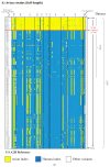
Heat map depicting timeline of adaptation to avian-to-human substitution (A2H) for all proteins of influenza A among publicly reported, full-length avian and human influenza A(H7N9) virus strains. Using the 109 A2H substitution sites identified from the 2014 dataset and the two new sites from 2017 dataset as a reference, corresponding signature residues for each reported avian (A) and human (B) H7N9 strain sequence are shown in alignment to the reference and arranged in chronological order of strain isolation (up to 2016 for avian strains and 2015 for human strains; only strains reported with full-length protein sequences were analyzed; full-length strains that could not be ascertained by the accession were ignored). The signature columns within each protein show the residue observed at each of the A2H substitution sites. Each strain is annotated with subtype, year and country of isolation, and isolate name. The first and the last pattern of the alignment are the avian-to-human substitution (A2H) residues, with the avian index sequence as the first (top) pattern and the human index sequence as the last pattern (bottom). Signature residues characteristic of the avian index are shown on a yellow background, while residues characteristic of the human index are shown on a dark blue background, and all other variants are on white. A higher resolution of the image, with visible details, is provided in Supplementary Table S7.
Figure 5
Figure 5
Heat map depicting timeline of adaptation to avian-to-human substitution (A2H) for all proteins of influenza A among publicly reported, full-length avian and human influenza A(H7N9) virus strains. Using the 109 A2H substitution sites identified from the 2014 dataset and the two new sites from 2017 dataset as a reference, corresponding signature residues for each reported avian (A) and human (B) H7N9 strain sequence are shown in alignment to the reference and arranged in chronological order of strain isolation (up to 2016 for avian strains and 2015 for human strains; only strains reported with full-length protein sequences were analyzed; full-length strains that could not be ascertained by the accession were ignored). The signature columns within each protein show the residue observed at each of the A2H substitution sites. Each strain is annotated with subtype, year and country of isolation, and isolate name. The first and the last pattern of the alignment are the avian-to-human substitution (A2H) residues, with the avian index sequence as the first (top) pattern and the human index sequence as the last pattern (bottom). Signature residues characteristic of the avian index are shown on a yellow background, while residues characteristic of the human index are shown on a dark blue background, and all other variants are on white. A higher resolution of the image, with visible details, is provided in Supplementary Table S7.

Similar articles

Cited by

References

    1. Bouvier N.M., Palese P. The biology of influenza viruses. Vaccine. 2008;26:D49–D53. doi: 10.1016/j.vaccine.2008.07.039. - DOI - PMC - PubMed
    1. Taubenberger J.K., Kash J.C. Influenza virus evolution, host adaptation, and pandemic formation. Cell Host Microbe. 2010;7:440–451. doi: 10.1016/j.chom.2010.05.009. - DOI - PMC - PubMed
    1. Nowak M.A. What is a quasispecies? Trends Ecol. Evol. 1992;7:118–121. doi: 10.1016/0169-5347(92)90145-2. - DOI - PubMed
    1. Steinhauer D.A. Influenza: Pathways to human adaptation. Nature. 2013;499:412–413. doi: 10.1038/nature12455. - DOI - PubMed
    1. Fitch W.M., Leiter J.M., Li X.Q., Palese P. Positive Darwinian evolution in human influenza A viruses. Proc. Natl. Acad. Sci. USA. 1991;88:4270–4274. doi: 10.1073/pnas.88.10.4270. - DOI - PMC - PubMed

Publication types