. 2021 May 10;22(9):5025.
doi: 10.3390/ijms22095025.

Dystrophin Deficiency Causes Progressive Depletion of Cardiovascular Progenitor Cells in the Heart

Affiliations

Dystrophin Deficiency Causes Progressive Depletion of Cardiovascular Progenitor Cells in the Heart

Sarka Jelinkova et al. Int J Mol Sci. .

Abstract

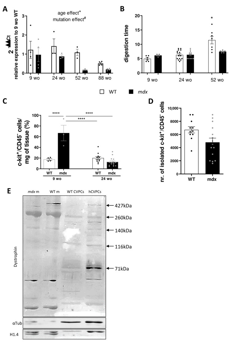
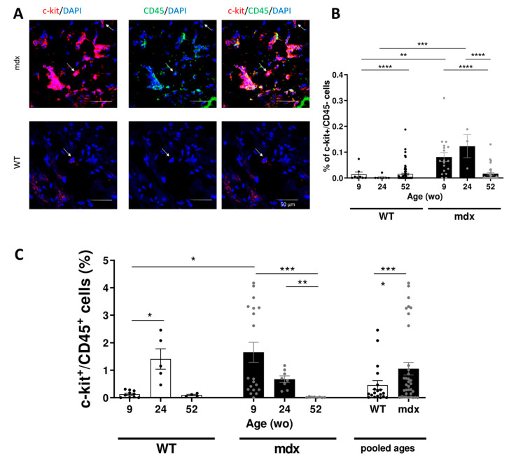
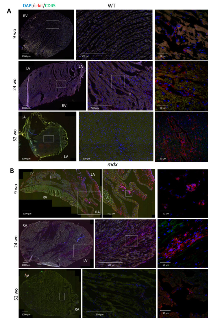
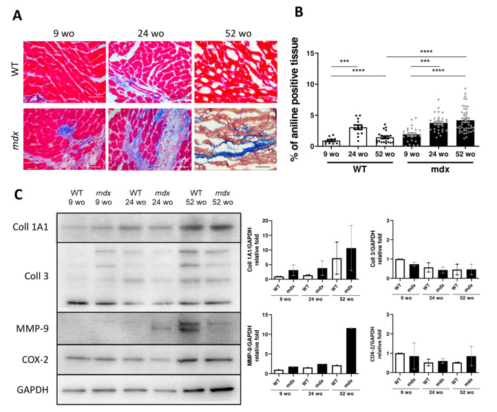
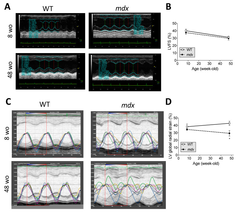
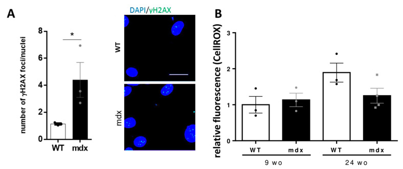
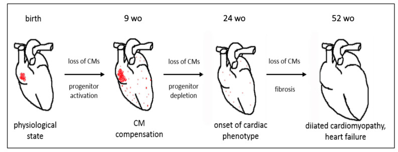
Duchenne muscular dystrophy (DMD) is a devastating condition shortening the lifespan of young men. DMD patients suffer from age-related dilated cardiomyopathy (DCM) that leads to heart failure. Several molecular mechanisms leading to cardiomyocyte death in DMD have been described. However, the pathological progression of DMD-associated DCM remains unclear. In skeletal muscle, a dramatic decrease in stem cells, so-called satellite cells, has been shown in DMD patients. Whether similar dysfunction occurs with cardiac muscle cardiovascular progenitor cells (CVPCs) in DMD remains to be explored. We hypothesized that the number of CVPCs decreases in the dystrophin-deficient heart with age and disease state, contributing to DCM progression. We used the dystrophin-deficient mouse model (mdx) to investigate age-dependent CVPC properties. Using quantitative PCR, flow cytometry, speckle tracking echocardiography, and immunofluorescence, we revealed that young mdx mice exhibit elevated CVPCs. We observed a rapid age-related CVPC depletion, coinciding with the progressive onset of cardiac dysfunction. Moreover, mdx CVPCs displayed increased DNA damage, suggesting impaired cardiac muscle homeostasis. Overall, our results identify the early recruitment of CVPCs in dystrophic hearts and their fast depletion with ageing. This latter depletion may participate in the fibrosis development and the acceleration onset of the cardiomyopathy.

Keywords: c-kit; cardiovascular progenitors; dilated cardiomyopathy; duchenne muscular dystrophy; genomic instability; mdx mouse.


Conflict of interest statement

The authors declare that the research was conducted in the absence of any commercial or financial relationships that could be construed as a potential conflict of interest.

Figures

Figure 1
Figure 1
C-kit expression decreases in mdx animals in an age-dependent manner. (A) Bar graphs summarizing the relative c-KIT gene expression analyzed by RT-PCR in WT (open bars and black dots) and mdx (black bars and grey dots) cardiac left ventricles. Data are shown as mean ± SEM (n = 3–4 animals in each group). The presented value was calculated as 2-ΔΔCt compared to housekeeping gene and 9 wo WT animals. Statistical difference was calculated by two-way ANOVA with Tukey’s post-hoc test with mutation (# p < 0.05 for WT compared to mdx) and age (* p < 0.05 for mdx ageing mouse) as evaluation criteria. (B) Bar graphs summarizing the digestion time (in minutes) to dissociate the mouse heart. Data are shown as mean ± SEM (n = 5–15 animals in each group). Kruskal–Wallis test with Dunn’s multiple comparison was used for statistical evaluation (* p < 0.05). (C) Ratio of c-kit+/CD45 cells isolated by MACS after cardiomyocyte filtration from 9 wo and 24 wo WT (open bars and black dots, n = 4–11) and mdx (black bars and grey dots, n = 3–16) mice, normalized by heart weight. Data are shown as mean ± SEM. Statistical significance was calculated using two-way ANOVA with Tukey’s post-hoc test (**** p < 0.0001). (D) Number of c-kit+/CD45 cells isolated by MACS after cardiomyocyte filtration in 24 wo WT mice (open bars and black dots, n = 11 animals) and 24 wo mdx mice (black bars and grey dots, n = 15 animals). (E) Illustrative images of Western blot (n = 3 repetitions) to evaluate the dystrophin expression in mouse (WT CVPCs) and human c-kit+ (hCVPCs) isolated cells. The WT mouse skeletal muscle tissue (WT m) was used as positive control, while mdx skeletal muscle tissue (mdx m) was used to show no expression of the long isoform of dystrophin (Dp427) in mdx animal. The α-tubulin and basic histone H1.4 were used as loading controls.
Figure 2
Figure 2
Recruitment of CVPCs in mdx hearts. (A) Representative histological analysis images of c-kit and CD45 immunolabeling. CVPCs were evaluated only as cells with positive signal for c-kit (red) and without signal for CD45 (green). Individual cells with these criteria are identified by arrows on the images. C-kit+/CD45 cells were evaluated as mast cells. Scale bars: 50 μm. (B) Bar graphs showing the percentage of c-kit+/CD45 cells relative to the total number of cells in WT (open bar and black dots) and mdx (black bar and grey dots). Data are shown as mean ± SEM (n = 3–20 sections per group; three animals were analyzed for the 9 wo and 52 wo groups, and two animals were analyzed for the 24 wo group). The statistical significance was calculated by two-way ANOVA and Tukey’s multiple comparison test (* p < 0.05, ** p < 0.01, *** p < 0.001). (C) Bar graphs showing the percentage of inflammatory mast cells (c-kit+/CD45) at different ages in WT (white bars and black dots) and mdx (black bars and grey dots) hearts. These mast cells were evaluated from the labeled sections shown in Figure 2A. Data are shown as mean ± SEM. Statistical significance was calculated by Kruskal–Wallis test and Dunn’s multiple comparisons for each individual age group of WT and mdx hearts and by Student’s t-test for pooled age groups (* p < 0.05, ** p < 0.01, *** p < 0.001, **** p < 0.0001).
Figure 3
Figure 3
Abnormal age-dependent CVPC localization in the mdx heart. (A) Representative images of CVPC localization in WT hearts at the age of 9 (upper panels, n = 3 animals), 24 (middle panels, n = 2 animals), and 52 wo (lower panels, n = 4 animals). (B) Representative images of CVPC localization in mdx hearts at the age of 9 wo (upper panel, three animals), 24 wo (middle panel, two animals) and 52 wo (lower panel, four animals). RV: right ventricle, LV: left ventricle, RA: right atrium, LA: left atrium. The c-kit protein was labeled in red, CD45 protein in green, and the nuclear DNA was counterstained with DAPI in blue. The detailed CVPC image taken by confocal microscopy was supplemented to the overview of the slice taken on fluorescent microscope and exactly positioned in the figure relative to the overall slice (left panels taken by confocal microscope, right and middle panel show detailed section marked in white rectangle). For each image the scale bar is indicated.
Figure 4
Figure 4
Fibrotic deposit and cardiac dysfunction correlate with decreasing CVPC presence in mdx heart. (A) Representative images of histological analysis stained using Masson trichrome technique showing myocytes (in red) and collagenous fibrotic tissue (in blue) in the left ventricle of WT and mdx hearts at 9, 24, and 52 wo. Line represents 100 µm. (B) The ratio of red and blue stained tissue was evaluated in WT hearts (open bars and black dots, n = 4–11 slices/3 animals per group) and mdx hearts (black bars and grey dots, n = 3–16 slices/3 animals per group) at the age of 24 wo and further at 52 wo. Statistical significance was calculated by Kruskal–Wallis test and Dunn‘s multiple comparison post-hoc test (*** p < 0.001, **** p < 0.0001). (C) Western blot analysis of collagen proteins and inflammatory proteins in the cardiac tissues. Left panel shows representative images of collagen 1A1 (Coll 1A1), collagen 3 (Coll 3), cyclooxygenase 2 (COX-2), and matrix metalloproteinase 9 (MMP-9) compared to the GAPDH control. The right panels show the normalized densitometry of each protein normalized by GAPDH content of WT (open bars, n = 2 animals) and mdx (black bars, n = 2 animals).
Figure 5
Figure 5
LV global strains are altered before LV ejection fraction in mdx mice. (A) M-mode images in WT and mdx at 8 and 48 wo. (B) Percentage (%) of the left ventricular fractional shortening (LVFS) in WT and mdx mice from 8 to 48 wo. (C) High-frequency ultrasound-based two-dimensional speckle-tracking (STE) images in WT and mdx at 8 and 48 wo. (D) Percentage (%) of the left ventricular (LV) global radial global strain in WT and mdx mice from 8 to 48 wo. Data are shown as mean ± SEM (n = 10 animals/group). A two-way ANOVA test was performed. * p < 0.05, mdx vs. WT mice at the same age.
Figure 6
Figure 6
DNA damage is increased in mdx CVPCs. (A) Bar graphs showing the DNA double stranded breaks in isolated CVPCs from 24 wo WT and mdx mice as illustrated with the two images on the right. The DNA damage was evaluated as the ratio of γH2AX foci number (in green) per total number of nuclei (DAPI labeled, in blue) per image. At least 20 cells were evaluated from each animal; the average from each animal was used for statistics with three animals per group (* p < 0.05). The scale bar represents 20 µm. (B) Bar graphs showing the relative fluorescence of ROS production using CellROX dye on freshly isolated CVPCs of 9 and 24 wo mice. At least three animals per group were used. The values are represented in the bar graphs for WT (open bars and black circles/dots) and mdx (black bars and grey dots). The statistical difference was calculated using two-way ANOVA.
Figure 7
Figure 7
Diagram of the proposed mechanism for the fate of CVPCs in the dystrophin-deficient heart. Dystrophin deficiency induces CVPC proliferation and hyperplasia in 9 wo mdx mice associated with an age-dependent depletion and simultaneous progressive fibrosis of the heart during ageing leading to development of cardiac dysfunction by 52 wo.

Similar articles

Cited by

References

    1. Mah J.K., Korngut L., Dykeman J., Day L., Pringsheim T., Jette N. A Systematic Review and Meta-Analysis on the Epidemiology of Duchenne and Becker Muscular Dystrophy. Neuromuscul. Disord. 2014;24:482–491. doi: 10.1016/j.nmd.2014.03.008. - DOI - PubMed
    1. Nigro G., Comi L.I., Politano L., Bain R.J. The Incidence and Evolution of Cardiomyopathy in Duchenne Muscular Dystrophy. Int. J. Cardiol. 1990;26:271–277. doi: 10.1016/0167-5273(90)90082-G. - DOI - PubMed
    1. Clarac F., Massion J., Smith A.M. Duchenne, Charcot and Babinski, Three Neurologists of La Salpetrière Hospital, and Their Contribution to Concepts of the Central Organization of Motor Synergy. J. Physiol. 2009;103:361–376. doi: 10.1016/j.jphysparis.2009.09.001. - DOI - PubMed
    1. Amedro P., Vincenti M., De La Villeon G., Lavastre K., Barrea C., Guillaumont S., Bredy C., Gamon L., Meli A.C., Cazorla O., et al. Speckle-Tracking Echocardiography in Children with Duchenne Muscular Dystrophy: A Prospective Multicenter Controlled Cross-Sectional Study. J. Am. Soc. Echocardiogr. 2019;32:412–422. doi: 10.1016/j.echo.2018.10.017. - DOI - PubMed
    1. Manzur A.Y., Kinali M., Muntoni F. Update on the Management of Duchenne Muscular Dystrophy. Arch. Dis. Child. 2008;93:986–990. doi: 10.1136/adc.2007.118141. - DOI - PubMed

MeSH terms

LinkOut - more resources