. 2021 May 10;11(5):427.
doi: 10.3390/life11050427.

Natural Sulfurs Inhibit LPS-Induced Inflammatory Responses through NF-κB Signaling in CCD-986Sk Skin Fibroblasts

Affiliations

Natural Sulfurs Inhibit LPS-Induced Inflammatory Responses through NF-κB Signaling in CCD-986Sk Skin Fibroblasts

Nipin Sp et al. Life (Basel). .

Abstract

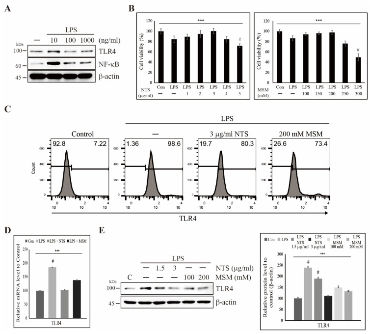
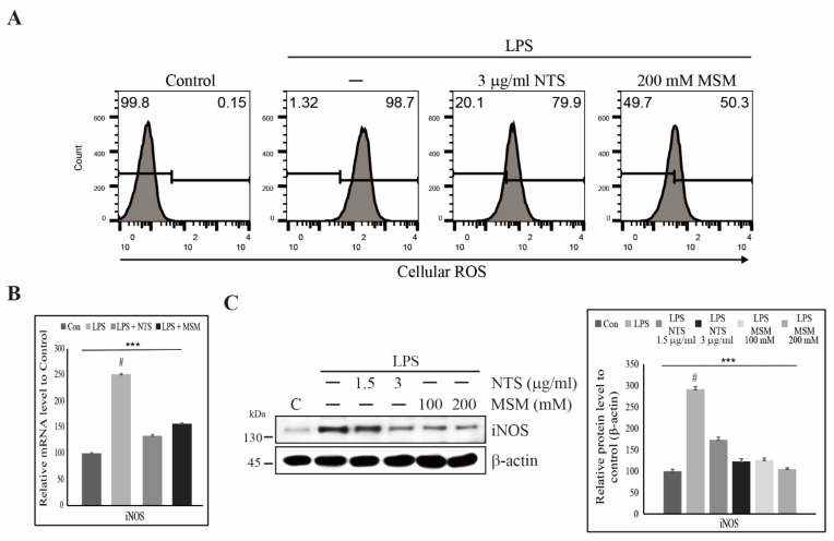
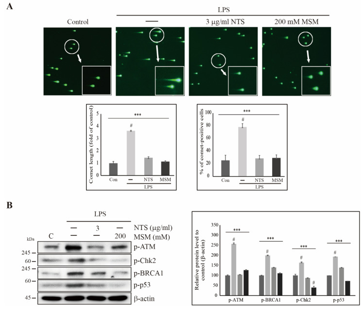
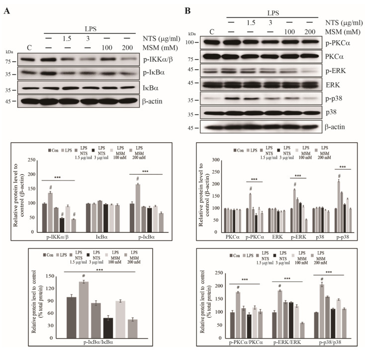
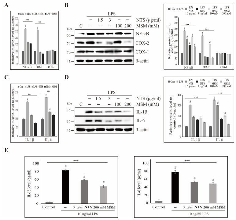
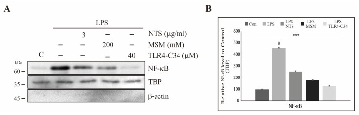
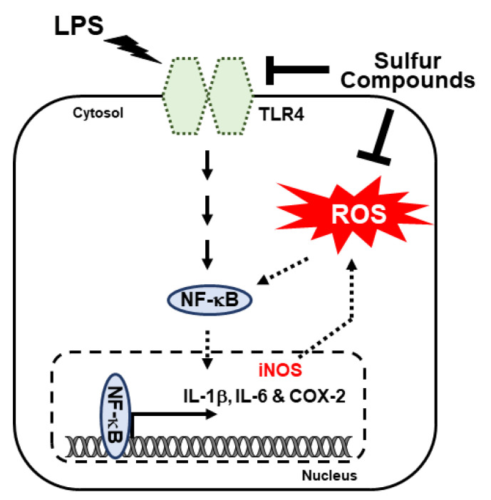
Lipopolysaccharide (LPS)-induced inflammatory response leads to serious damage, up to and including tumorigenesis. Natural mineral sulfur, non-toxic sulfur (NTS), and methylsulfonylmethane (MSM) have anti-inflammatory activity that may inhibit LPS-induced inflammation. We hypothesized that sulfur compounds could inhibit LPS-induced inflammatory responses in CCD-986Sk skin fibroblasts. We used Western blotting and real-time PCR to analyze molecular signaling in treated and untreated cultures. We also used flow cytometry for cell surface receptor analysis, comet assays to evaluate DNA damage, and ELISA-based cytokine detection. LPS induced TLR4 activation and NF-κB signaling via canonical and protein kinase C (PKC)-dependent pathways, while NTS and MSM downregulated that response. NTS and MSM also inhibited LPS-induced nuclear accumulation and binding of NF-κB to proinflammatory cytokines COX-2, IL-1β, and IL-6. Finally, the sulfur compounds suppressed LPS-induced ROS accumulation and DNA damage in CCD-986Sk cells. These results suggest that natural sulfur compounds could be used to treat inflammation and may be useful in the development of cosmetics.

Keywords: CCD-986Sk; LPS; MSM; NF-κB; NTS; ROS; TLR4; anti-inflammatory effect.


Conflict of interest statement

The authors have no conflict of interest to declare.

Figures

Figure 1
Figure 1
Sulfur compounds inhibited LPS-induced expression of TLR4. (A) Western blot analysis in CCD-986Sk cells treated with 10, 100, and 1000 ng/mL LPS for 48 h showing TLR4 and NF-κB expression. (B) MTT assay showing cell viability with 10 ng/mL LPS and increasing concentrations of NTS or MSM. Data are representative of three independent experiments. *** p < 0.001 (ANOVA test). # The mean difference is significant at the 0.001 level. (C) Flow cytometry showing inhibition of LPS-induced expression of TLR4 by 3 µg/mL NTS and 200 mM MSM for 48 h. (D) Real-time PCR data of mRNA after treatment with 10 ng/mL LPS and 3 µg/mL NTS or 200 mMMSM for 48 h showing the relative expression of TLR4 normalized to GAPDH. *** p < 0.001 (ANOVA test). # The mean difference is significant at the 0.001 level. (E) Western blotting analysis of TLR4 expression in CCD-986Sk cells treated with 10 ng/mL LPS and NTS (1.5 and 3 ng/mL) or MSM (100 and 200 mM) for 48 h. Expression levels were determined by densitometry and normalized to β-actin. Data are representative of three independent experiments. *** p < 0.001 (ANOVA test). # The mean difference is significant at the 0.001 level.
Figure 2
Figure 2
Sulfur compounds inhibited LPS-induced ROS. (A) Flow cytometry analysis of cellular ROS in CCD-986Sk cells after treatment with 10 ng/mL LPS and 3 µg/mL NTS or 200 mM MSM for 48 h. (B) Real-time PCR of iNOS mRNA after treatment with 10 ng/mL LPS and 3 µg/mL NTS or 200 mM MSM after 48 h, data normalized to GAPDH. *** p < 0.001 (ANOVA test). # The mean difference was significant at the 0.001 level. (C) Western blotting analysis of iNOS expression in CCD-986Sk cells treated with 10 ng/mL LPS and NTS (1.5 and 3 ng/mL) or MSM (100 and 200 mM) for 48 h. The representative expression levels of proteins following NTS or MSM treatment were determined by densitometry and normalized to β-actin. Data are representative of three independent experiments. *** p < 0.001 (ANOVA test). # The mean difference was significant at the 0.001 level.
Figure 3
Figure 3
Sulfur compounds inhibited LPS-induced DNA damage. (A) Comet assay imaged by electron microscopy for 10× and 40× magnification showing fragmented DNA migrated out of the nucleoid body and formed a comet tail after treatment with 10 ng/mL LPS, 3 µg/mL NTS, or 200 mM MSM for 48 h. Representative comet length and comet-positive cells were analyzed as the fold change versus the control. Statistical analysis was performed using the ANOVA test (*** p < 0.001). # Mean difference was significant at the 0.001 level. (B) Western blot analysis in CCD-986Sk cells showing that 3 µg/mL of NTS or 200 mM of MSM inhibited LPS-induced expressions of p-ATM, p-Chk2, p-BRCA1, and p-p53. Expression was determined by densitometry and normalized to β-actin. The values are presented as means ± SEM of three independent experiments performed in duplicate (n = 3). *** p < 0.001 (ANOVA test). # Mean difference was significant at the 0.001 level.
Figure 4
Figure 4
Sulfur compounds inhibited the LPS-induced canonical and PKC-dependent NF-κB pathways. (A) Western blotting analysis in CCD-986Sk cells treated with 1.5 and 3 µg/mL of NTS or 100 and 200 mM of MSM for 48 h showing the inhibition of 10 ng/mL LPS-induced p-IKKα/β, IκBα, and p-IκBα expression. Protein expression was determined by densitometry and normalized to β-actin. Values are presented as means ± SEM of three independent experiments performed in duplicate (n = 3). *** p < 0.001 (ANOVA test). Relative expressions of p-IκBα protein were determined via densitometry and normalized to total IκBα protein. *** p < 0.001. # Mean difference was significant at the 0.001 level. (B) Western blotting analysis in CCD-986Sk cells treated with 1.5 and 3 µg/mL of NTS or 100 and 200 mM of MSM for 48 h showing inhibition of 10 ng/mL LPS-induced p-PKC-α, p-ERK, and p-p38 expression along with unchanged expressions of PKC-α, ERK, and p38 proteins. Protein expression was determined by densitometry and normalized to β-actin. The values are presented as means ± SEM of three independent experiments performed in duplicate (n = 3). *** p < 0.001 (ANOVA test). Relative expressions of p-PKC-α, p-ERK, and p-p38 were determined via densitometry and normalized to their total PKC-α, ERK, and p38 proteins. *** p < 0.001. # Mean difference was significant at the 0.001 level.
Figure 5
Figure 5
Sulfur compounds inhibited the LPS-induced NF-κB, COX-2, and proinflammatory cytokines. (A) Real-time PCR data of mRNA after treatment with 10 ng/mL LPS and 3 µg/mL NTS and 200 mM MSM for 48 h showing the relative expression levels of NF-κB, COX-1, and COX-2 normalized to GAPDH mRNA. *** p < 0.001 (ANOVA test). # The mean difference was significant at the 0.001 level. (B) Western blotting analysis in CCD-986Sk cells treated with 10 ng/mL LPS and NTS (1.5 and 3 ng/mL) or MSM (100 and 200 mM) for 48 h showing the expression of NF-κB, COX-1, and COX-2. The representative expression levels of proteins following NTS or MSM treatment were determined by densitometry and normalized to β-actin. Data are representative of three independent experiments. *** p < 0.001 (ANOVA test). # The mean difference was significant at the 0.001 level. (C) Real-time PCR data of mRNA after treatment with 10 ng/mL LPS and 3 µg/mL of NTS and 200 mM of MSM for 48 h showing the relative expression levels of IL-1β and IL-6 normalized to GAPDH mRNA. *** p < 0.001 (ANOVA test). # The mean difference was significant at the 0.001 level. (D) Western blotting analysis in CCD-986Sk cells treated with 10 ng/mL LPS and NTS (1.5 and 3 ng/mL) or MSM (100 and 200 mM) for 48 h showing the expression of IL-1β and IL-6. The representative expression levels of proteins following NTS or MSM treatment were determined by densitometry and normalized to β-actin. Data are representative of three independent experiments. *** p < 0.001 (ANOVA test). # The mean difference was significant at the 0.001 level. (E) ELISA analysis showing the inhibition of the LPS-induced expression of IL-1β and IL-6 by 3 µg/mL of NTS and 200 mM of MSM. Data are representative of three independent experiments. *** p < 0.001 (ANOVA test). # The mean difference was significant at the 0.001 level.
Figure 6
Figure 6
Sulfur compounds inhibited the LPS-induced nuclear accumulation of NF-κB. (A) Western blotting analysis of nuclear extracts harvested from CCD-986Sk cells treated with 3 µg/mL NTS or 200 mM MSM or 40 µM TLR4 inhibitor (TLR4-C34) for 48 h showing the inhibition of 10 ng/mL LPS-induced nuclear translocation of NF-κB. (B) Protein expression after NTS, MSM, and TLR4-C34 treatment were determined by densitometry and normalized to TBP. Data are representative of three independent experiments. *** p < 0.001 (ANOVA test). # The mean difference was significant at the 0.001 level.
Figure 7
Figure 7
Molecular mechanism of LPS-induced regulation of the inflammatory response by TLR4/NF-κB through canonical and PKC-mediated pathways. The anti-inflammatory activities of NTS and MSM were achieved by inhibiting these pathways and binding of NF-κB to proinflammatory cytokines.

Similar articles

Cited by

References

    1. Gvirtz R., Ogen-Shtern N., Cohen G. Kinetic cytokine secretion profile of lps-induced inflammation in the human skin organ culture. Pharmaceutics. 2020;12:299. doi: 10.3390/pharmaceutics12040299. - DOI - PMC - PubMed
    1. Gonzales K.A.U., Fuchs E. Skin and its regenerative powers: An alliance between stem cells and their niche. Dev. Cell. 2017;43:387–401. doi: 10.1016/j.devcel.2017.10.001. - DOI - PMC - PubMed
    1. Jordana M., Sarnstrand B., Sime P.J., Ramis I. Immune-inflammatory functions of fibroblasts. Eur. Respir. J. 1994;7:2212–2222. doi: 10.1183/09031936.94.07122212. - DOI - PubMed
    1. Palm E., Khalaf H., Bengtsson T. Porphyromonas gingivalis downregulates the immune response of fibroblasts. BMC Microbiol. 2013;13:155. doi: 10.1186/1471-2180-13-155. - DOI - PMC - PubMed
    1. Xi X., McMillan D.H., Lehmann G.M., Sime P.J., Libby R.T., Huxlin K.R., Feldon S.E., Phipps R.P. Ocular fibroblast diversity: Implications for inflammation and ocular wound healing. Invest. Ophthalmol. Vis. Sci. 2011;52:4859–4865. doi: 10.1167/iovs.10-7066. - DOI - PMC - PubMed