. 2021 May 10;10(5):1151.
doi: 10.3390/cells10051151.

CD47 Potentiates Inflammatory Response in Systemic Lupus Erythematosus

Affiliations

CD47 Potentiates Inflammatory Response in Systemic Lupus Erythematosus

Jin Kyun Park et al. Cells. .

Abstract

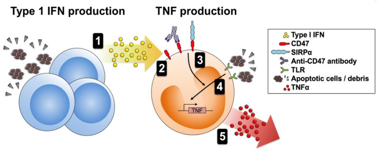
Background: To investigate the role of CD47 in inflammatory responses in systemic lupus erythematosus (SLE).

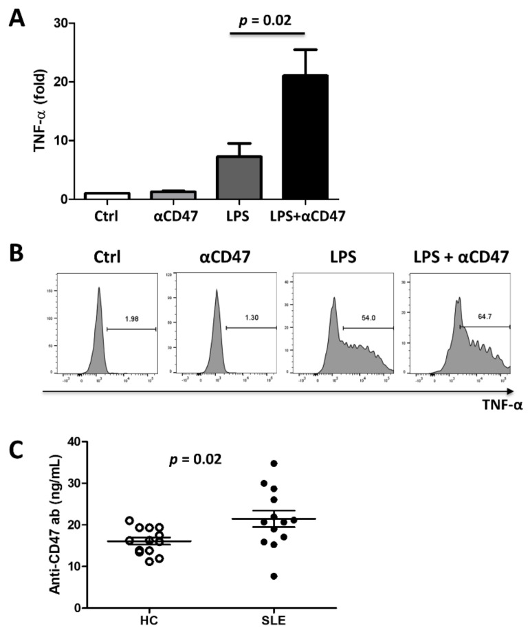
Methods: Expression of CD47 and signal regulatory protein alpha (SIRPα) by peripheral blood mononuclear cells (PBMCs) and changes in CD47 expression after exposure to SLE serum, healthy control (HC) serum, recombinant interferon (IFN)-α, or tumor necrosis factor (TNF)-α were examined. Human monocytes and THP1 cells were incubated with lipopolysaccharide (LPS), an anti-CD47 antibody, or both. TNF-α production was examined. Sera from SLE patients and HCs were screened to detect autoantibodies specific for CD47.

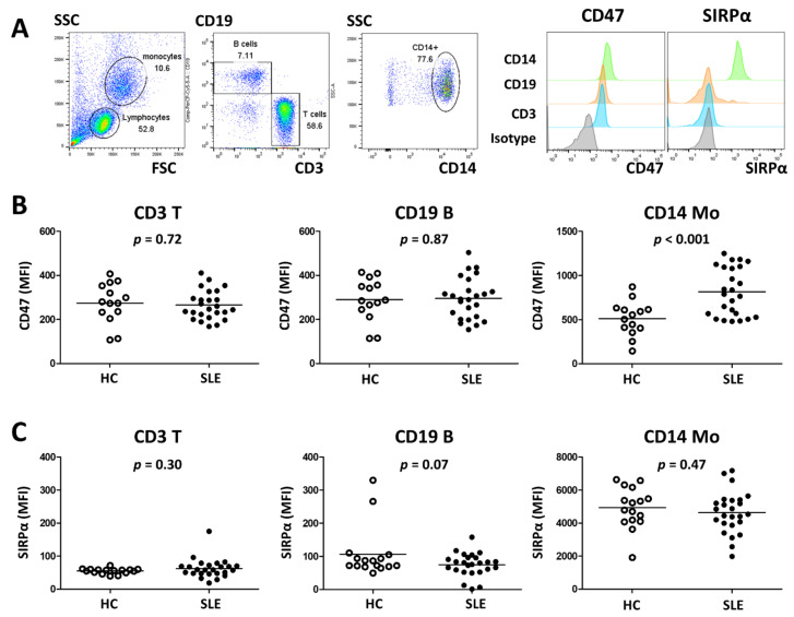
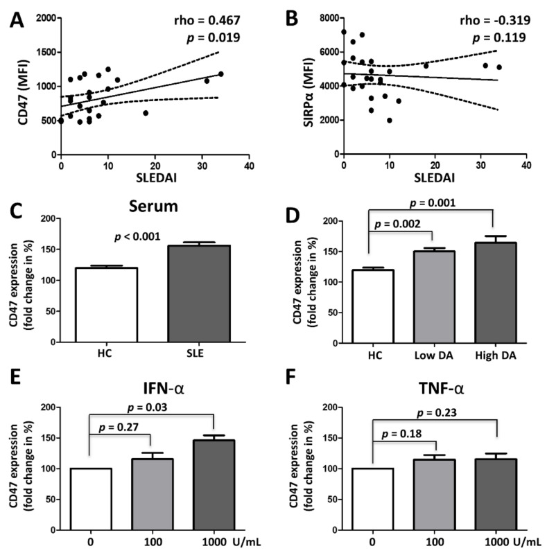
Results: Twenty-five SLE patients and sixteen HCs were enrolled. CD47 expression by monocytes from SLE patients was higher than those from HCs (mean fluorescence intensity ± SD: 815.9 ± 269.4 vs. 511.5 ± 199.4, respectively; p < 0.001). CD47 expression by monocytes correlated with SLE disease activity (Spearman's rho = 0.467, p = 0.019). IFN-α but not TNF-α, increased CD47 expression. Exposing monocytes to an anti-CD47 antibody plus LPS increased TNF-α production by 21.0 ± 10.9-fold (compared with 7.3 ± 5.5-fold for LPS alone). Finally, levels of autoantibodies against CD47 were higher in SLE patients than in HCs (21.4 ± 7.1 ng/mL vs. 16.1 ± 3.1 ng/mL, respectively; p = 0.02). Anti-CD47 antibody levels did not correlate with disease activity (Spearman's rho = -0.11, p = 0.759) or CD47 expression on CD14 monocytes (Spearman's rho = 0.079, p = 0.838) in patients.

Conclusions: CD47 expression by monocytes is upregulated in SLE and correlates with disease activity. CD47 contributes to augmented inflammatory responses in SLE. Targeting CD47 might be a novel treatment for SLE.

Keywords: CD47; SIRP-alpha; inflammatory response; systemic lupus erythematosus.


Conflict of interest statement

The authors declare that they have no competing interests.

Figures

Figure 1
Figure 1
CD47 expression by SLE monocytes is upregulated. (A) PBMCs from 25 SLE patients and 14 healthy controls (HCs) were examined with respect to CD47 and SIRPα expression by flow cytometry. CD19+ B cells, CD3+ T cells, and CD14+ monocytes were gated (left panel) and representative surface expression of CD47 and SIRPα were depicted (right panel). CD47 expression (B) and SIRPα expression (C) on T cells, B cells and monocytes were compared between HC and SLE. p values were generated using t-tests. MFI, mean fluorescence intensity; Mo, monocytes.
Figure 2
Figure 2
Upregulation of CD47 by SLE serum or inflammatory cytokines. Expression of CD47 (A) and SIRPα (B) on monocytes from SLE patients were correlated with their disease activity. p values were generated by Spearman’s correlation analysis. (C and D) Healthy PBMCs were incubated with serum from healthy controls (n = 6) and SLE patients (n = 10), and fold changes in CD47 expression on monocytes were investigated by flow cytometry analysis. (D) Effect of serum from patients with low (n = 6) and high (n = 4) disease activity on CD47 expression was examined. High and low disease activity were defined as SLEDAI > 12 or SLEDAI <12, respectively. (E and F) Healthy PBMCs (n = 3) were incubated with increasing concentrations of interferon-alpha (IFN-α) and tumor necrosis factor-alpha (TNF-α) and change in CD47 expression was examined by flow cytometry. Untreated samples served as a reference (i.e., 100%). p values were generated using t-tests. DA, disease activity; MFI, mean fluorescence intensity; SLE, systemic lupus erythematosus; SLEDAI, SLE disease activity index 2000.
Figure 3
Figure 3
CD47 activation potentiates inflammatory responses. (A) PBMCs from healthy controls (n = 6) were stimulated for 5 hours with a mouse anti-human CD47 antibody, LPS (3 ng/mL), or both, and TNF-α production in CD14+ monocytes was measured by flow cytometry. Treatment with an anti-human CD47 antibody and LPS induced a significantly greater increase in TNF-α production in monocytes than treatment with LPS alone. (B) Representative flow cytometry plots are depicted. (C) Serum from 13 SLE patients and 13 healthy controls (HCs) was screened for anti-CD47 antibodies using ELISA. Serum anti-CD47 antibody levels were significantly higher in SLE patients than in HCs. p value was generated using a t-test. αCD47, anti-CD47 mouse monoclonal antibody; Ctrl, control; HC, health controls; LPS, lipopolysaccharide; SLE, systemic lupus erythematosus; TNF, tumor necrosis factor.
Figure 4
Figure 4
Presence of CD47 expressing macrophages in lupus nephritis. (A) Kidney sections from patients with lupus nephritis (LN, n = 7) were stained with hematoxylin and eosin (H&E) (magnification 40×). Tissue sections were stained with DAPI (blue), CD47 (AF488, green), CD14 (AF647, magenta) and CD68 (AF594, red) by immunofluorescence. Representative images are shown. Arrows indicate co-expression of CD47 and CD68. Scale bar = 100 µm. (B) CD47 expressing CD68 cells (number/mm2) in LN according to the lupus nephritis class were counted. Data are mean ± SEM.
Figure 5
Figure 5
Proposed role for CD47 in SLE. (1) Immune and non-immune cells produce type 1 IFN when they encounter stimuli such as nucleic acids released from apoptotic debris. (2) Type 1 IFN increases CD47 expression by monocytes. (3) CD47 is activated by circulating autoantibodies; it then activates MAPK signaling components (including ERK, JNK, and p38). (4) LPS or other ligands bind to TLRs and activate NF-κB signaling. (5) Activated MAPK promotes transcription (and stability) and translation of TNF-α mRNA, leading to enhanced production of TNF-α protein. TLR, Toll-like receptor; LPS, lipopolysaccharide; TNF, tumor necrosis factor; IFN, interferon.

Similar articles

Cited by

References

    1. Rahman A., Isenberg D.A. Systemic lupus erythematosus. N. Engl. J. Med. 2008;358:929–939. doi: 10.1056/NEJMra071297. - DOI - PubMed
    1. Liu Z., Davidson A. Taming lupus—A new understanding of pathogenesis is leading to clinical advances. Nat. Med. 2012;18:871–882. doi: 10.1038/nm.2752. - DOI - PMC - PubMed
    1. Fleischer S.J., Daridon C., Fleischer V., Lipsky P.E., Dorner T. Enhanced Tyrosine Phosphatase Activity Underlies Dysregulated B Cell Receptor Signaling and Promotes Survival of Human Lupus B Cells. Arthritis Rheumatol. 2016;68:1210–1221. doi: 10.1002/art.39559. - DOI - PubMed
    1. Cella M., Dohring C., Samaridis J., Dessing M., Brockhaus M., Lanzavecchia A., Colonna M. A novel inhibitory receptor (ILT3) expressed on monocytes, macrophages, and dendritic cells involved in antigen processing. J. Exp. Med. 1997;185:1743–1751. doi: 10.1084/jem.185.10.1743. - DOI - PMC - PubMed
    1. Lu H.K., Rentero C., Raftery M.J., Borges L., Bryant K., Tedla N. Leukocyte Ig-like receptor B4 (LILRB4) is a potent inhibitor of FcgammaRI-mediated monocyte activation via dephosphorylation of multiple kinases. J. Biol. Chem. 2009;284:34839–34848. doi: 10.1074/jbc.M109.035683. - DOI - PMC - PubMed

Publication types