

Wound Repair and Extremely Low Frequency-Electromagnetic Field: Insight from In Vitro Study and Potential Clinical Application
- PMID: 34068809
- PMCID: PMC8126245
- DOI: 10.3390/ijms22095037
Wound Repair and Extremely Low Frequency-Electromagnetic Field: Insight from In Vitro Study and Potential Clinical Application
Abstract
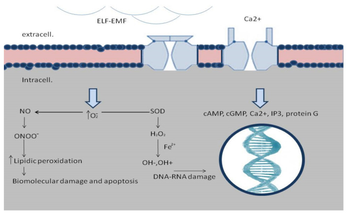
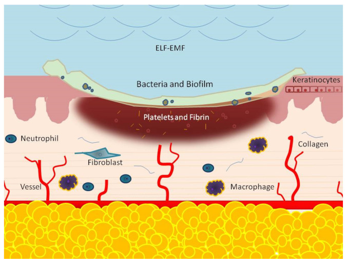
Wound healing is a complex, staged process. It involves extensive communication between the different cellular constituents of various compartments of the skin and its extracellular matrix (ECM). Different signaling pathways are determined by a mutual influence on each other, resulting in a dynamic and complex crosstalk. It consists of various dynamic processes including a series of overlapping phases: hemostasis, inflammation response, new tissue formation, and tissue remodeling. Interruption or deregulation of one or more of these phases may lead to non-healing (chronic) wounds. The most important factor among local and systemic exogenous factors leading to a chronic wound is infection with a biofilm presence. In the last few years, an increasing number of reports have evaluated the effects of extremely low frequency (ELF) electromagnetic fields (EMFs) on tissue repair. Each experimental result comes from a single element of this complex process. An interaction between ELF-EMFs and healing has shown to effectively modulate inflammation, protease matrix rearrangement, neo-angiogenesis, senescence, stem-cell proliferation, and epithelialization. These effects are strictly related to the time of exposure, waveform, frequency, and amplitude. In this review, we focus on the effect of ELF-EMFs on different wound healing phases.
Keywords: ELF-EMF; fibroblasts; healing; keratinocytes; non-healing wounds; wound.
Conflict of interest statement
All authors declare no conflict of interest.
Figures
Similar articles
-
Extremely low frequency electromagnetic field and wound healing: implication of cytokines as biological mediators.Eur Cytokine Netw. 2013 Mar;24(1):1-10. doi: 10.1684/ecn.2013.0332. Eur Cytokine Netw. 2013. PMID: 23674517 Review.
-
Extremely low-frequency electromagnetic fields accelerates wound healing modulating MMP-9 and inflammatory cytokines.Cell Prolif. 2018 Apr;51(2):e12432. doi: 10.1111/cpr.12432. Epub 2018 Jan 22. Cell Prolif. 2018. PMID: 29357406 Free PMC article.
-
Extremely low frequency electromagnetic fields modulate expression of inducible nitric oxide synthase, endothelial nitric oxide synthase and cyclooxygenase-2 in the human keratinocyte cell line HaCat: potential therapeutic effects in wound healing.Br J Dermatol. 2010 Feb 1;162(2):258-66. doi: 10.1111/j.1365-2133.2009.09527.x. Epub 2009 Oct 3. Br J Dermatol. 2010. PMID: 19799606
-
Human Gingival Fibroblasts Exposed to Extremely Low-Frequency Electromagnetic Fields: In Vitro Model of Wound-Healing Improvement.Int J Mol Sci. 2019 Apr 29;20(9):2108. doi: 10.3390/ijms20092108. Int J Mol Sci. 2019. PMID: 31035654 Free PMC article.
-
Trends in wound repair: cellular and molecular basis of regenerative therapy using electromagnetic fields.Curr Mol Med. 2012 Jan;12(1):14-26. doi: 10.2174/156652412798376143. Curr Mol Med. 2012. PMID: 22082478 Review.
Cited by
-
A Novel and Versatile Microfluidic Device for Cell Assays under Radio Frequency Exposure.Biosensors (Basel). 2023 Jul 27;13(8):763. doi: 10.3390/bios13080763. Biosensors (Basel). 2023. PMID: 37622849 Free PMC article.
-
Emerging cancer therapies: targeting physiological networks and cellular bioelectrical differences with non-thermal systemic electromagnetic fields in the human body - a comprehensive review.Front Netw Physiol. 2024 Dec 10;4:1483401. doi: 10.3389/fnetp.2024.1483401. eCollection 2024. Front Netw Physiol. 2024. PMID: 39720338 Free PMC article. Review.
-
The Cellular Response Is Determined by a Combination of Different ELF-EMF Exposure Parameters: A Scope Review.Int J Mol Sci. 2024 May 7;25(10):5074. doi: 10.3390/ijms25105074. Int J Mol Sci. 2024. PMID: 38791113 Free PMC article. Review.
-
Analgesic Effectiveness of Physical Therapy Combining the Use of Electromagnetic Fields with Light Radiation Emitted by LEDs along with the Use of Topical Herbal Ointment in Patients with Gonarthrosis.Int J Environ Res Public Health. 2023 Feb 19;20(4):3696. doi: 10.3390/ijerph20043696. Int J Environ Res Public Health. 2023. PMID: 36834396 Free PMC article.
-
[Research advances on the role of microRNA engineered exosomes in diabetic wounds].Zhonghua Shao Shang Yu Chuang Mian Xiu Fu Za Zhi. 2024 Feb 20;40(2):190-195. doi: 10.3760/cma.j.cn501225-20230721-00011. Zhonghua Shao Shang Yu Chuang Mian Xiu Fu Za Zhi. 2024. PMID: 38418181 Free PMC article. Review. Chinese.
References
Publication types
MeSH terms
LinkOut - more resources
Full Text Sources
Other Literature Sources
Medical
