Review
. 2021 May 14;10(10):2118.
doi: 10.3390/jcm10102118.

Rheumatoid Arthritis Associated Episcleritis and Scleritis: An Update on Treatment Perspectives

Affiliations
Review

Rheumatoid Arthritis Associated Episcleritis and Scleritis: An Update on Treatment Perspectives

Veronique Promelle et al. J Clin Med. .

Abstract

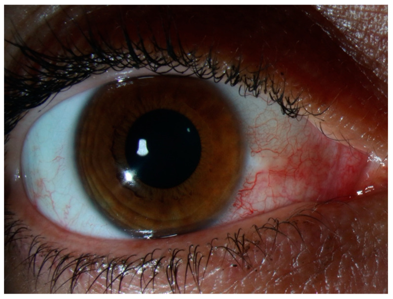
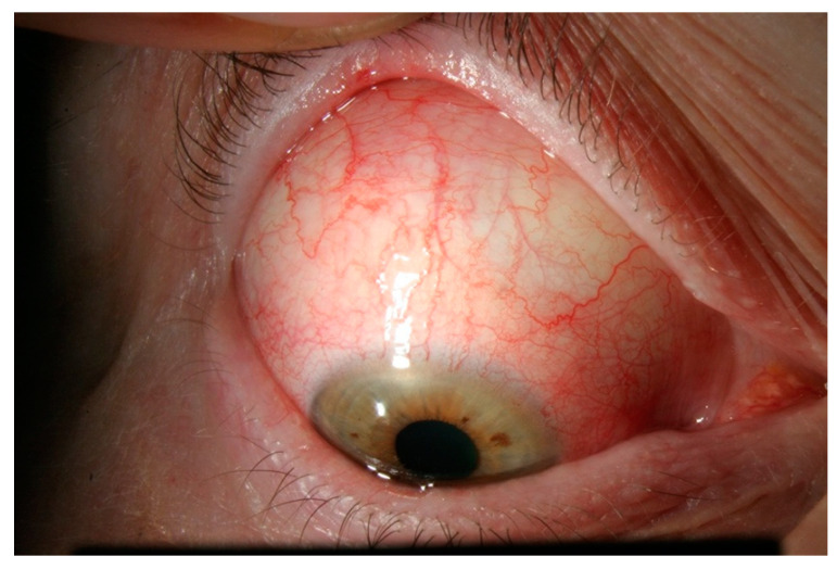
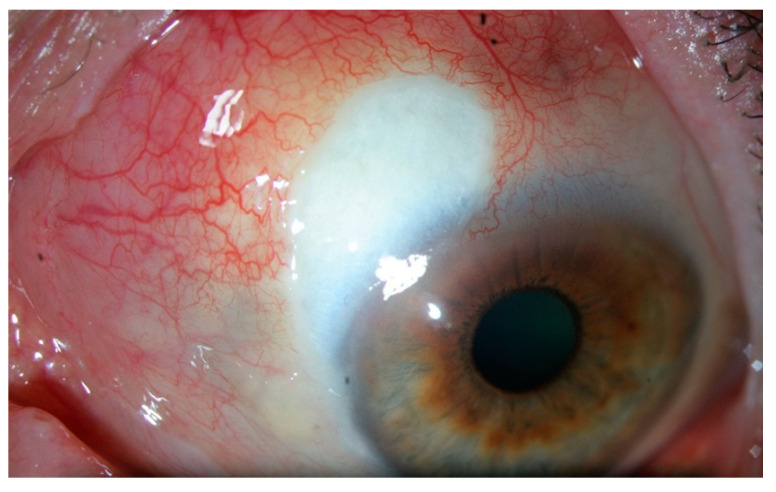
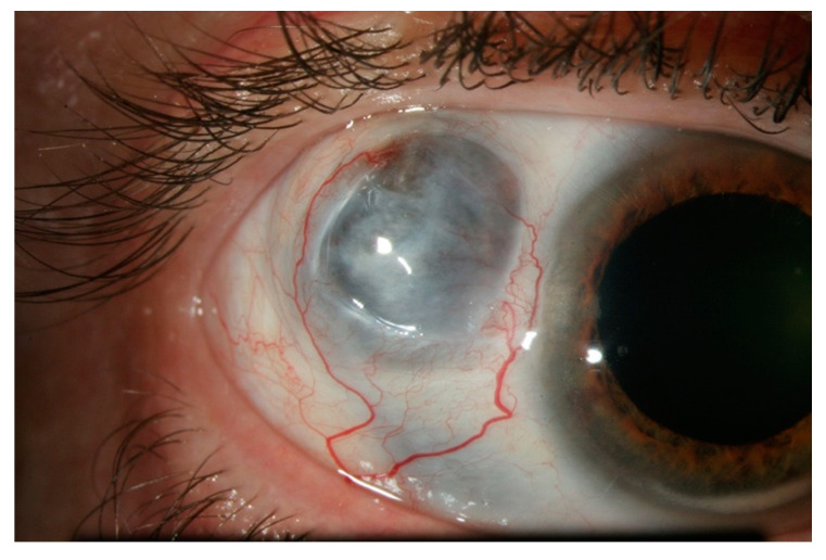
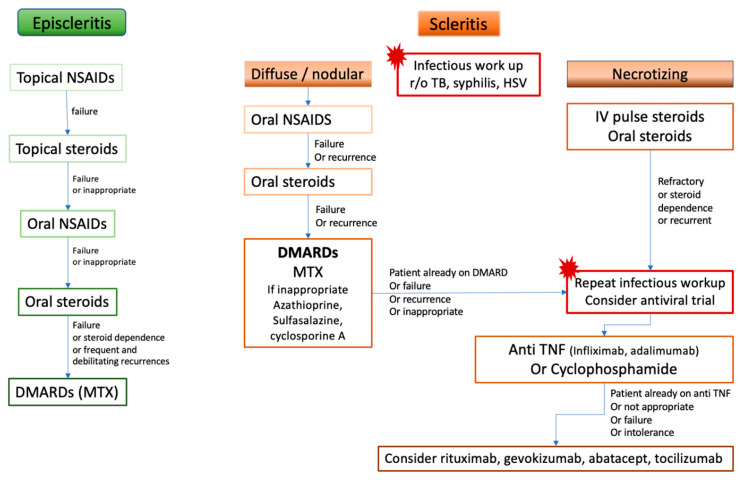
Episcleritis and scleritis are the most common ocular inflammatory manifestation of rheumatoid arthritis. Rheumatoid arthritis (RA) accounts for 8% to 15% of the cases of scleritis, and 2% of patients with RA will develop scleritis. These patients are more likely to present with diffuse or necrotizing forms of scleritis and have an increased risk of ocular complications and refractory scleral inflammation. In this review we provide an overview of diagnosis and management of rheumatoid arthritis-associated episcleritis and scleritis with a focus on recent treatment perspectives. Episcleritis is usually benign and treated with oral non-steroidal anti- inflammatory drugs (NSAIDs) and/or topical steroids. Treatment of scleritis will classically include oral NSAIDs and steroids but may require disease-modifying anti-rheumatic drugs (DMARDs). In refractory cases, treatment with anti TNF biologic agents (infliximab, and adalimumab) is now recommended. Evidence suggests that rituximab may be an effective option, and further studies are needed to investigate the potential role of gevokizumab, tocilizumab, abatacept, tofacitinib, or ACTH gel. A close cooperation is needed between the rheumatology or internal medicine specialist and the ophthalmologist, especially when scleritis may be the first indicator of an underlying rheumatoid vasculitis.

Keywords: biologics; episcleritis; ocular inflammation; prognosis; rheumatoid arthritis; scleritis; treatment.


Conflict of interest statement

The authors declare no conflict of interest.

Figures

Figure 1
Figure 1
Cross sectional diagram of the normal anatomy of the outer layers of the eye. The most superficial layer is the conjunctiva with the conjunctival vascular plexus, easily moved over the underlying structures. Underneath, the Tenon’s capsule and the episclera, with an episcleral plexus of straight, radially arranged vessels. The deepest is the scleral plexus, tightly adherent to the sclera.
Figure 2
Figure 2
Episcleritis. There is no scleral swelling, and although the deep vascular plexus is congested, oedema and infiltration are limited to the episclera. The eye redness is most often limited to one sector of the eye, with a salmon pink to red discoloration. The pain is generally mild.
Figure 3
Figure 3
Nodular anterior scleritis. A scleral nodule, totally immobile, lifts the vessels. The nodule is clearly separated from the overlying congested episcleral tissue.
Figure 4
Figure 4
Diffuse anterior scleritis. Widespread inflammation of sclera, and abnormal appearance of the deep scleral vascular plexus. The scleral plexus becomes visible after blanching the superficial vessels with phenylephrine.
Figure 5
Figure 5
Necrotizing scleritis with inflammation. This form presents like a diffuse scleritis with an area of avascular, necrotic sclera.
Figure 6
Figure 6
Necrotizing scleritis without inflammation, or scleromalacia perforans. This form presents with no inflammation and no pain, and almost complete destruction of scleral and episcleral tissue in one or more necrotic patch. The greyish appearance comes from the visible uveal tissue underneath.
Figure 7
Figure 7
Posterior scleritis. The scleral inflammation involves the posterior part of the globe, with no apparent external sign unless there is an associated anterior component. (A) B-scan ultrasound showing the classic T-sign from scleral oedema, (B) The scleral inflammation can cause exudative retinal detachment (*), retinal folds (dashed arrows), retinal veins congestion (arrows) and optic disc swelling.
Figure 8
Figure 8
Proposition of treatment algorithm and strategy of escalation in rheumatoid arthritis-associated episcleritis and scleritis. DMARDs: disease-modifying anti-rheumatic drugs, HSV: Herpes Simplex Virus, IV: intravenous, MTX: methotrexate, NSAIDs: non-steroidal anti-inflammatory drugs, r/o: rule out, TB: tuberculosis, TNF: Tumor necrosis factor.

Similar articles

Cited by

References

    1. Vignesh A.P.P., Srinivasan R. Ocular manifestations of rheumatoid arthritis and their correlation with anti-cyclic citrullinated peptide antibodies. Clin. Ophthalmol. 2015;9:393–397. doi: 10.2147/OPTH.S77210. - DOI - PMC - PubMed
    1. Itty S., Pulido J.S., Bakri S.J., Baratz K.H., Matteson E.L., Hodge D.O. Anti-Cyclic Citrullinated Peptide, Rheumatoid Factor, and Ocular Symptoms Typical of Rheumatoid Arthritis. Trans. Am. Ophthalmol. Soc. 2008;106:75–81. - PMC - PubMed
    1. Caimmi C., Crowson C.S., Smith W.M., Matteson E.L., Makol A. Clinical Correlates, Outcomes, and Predictors of Inflammatory Ocular Disease Associated with Rheumatoid Arthritis in the Biologic Era. J. Rheumatol. 2018;45:595–603. doi: 10.3899/jrheum.170437. - DOI - PubMed
    1. Bettero R.G., Cebrian R.F.M., Skare T.L. Prevalence of ocular manifestation in 198 patients with rheumatoid arthritis: A retrospective study. Arq. Bras. Oftalmol. 2008;71:365–369. doi: 10.1590/S0004-27492008000300011. - DOI - PubMed
    1. Zlatanović G., Veselinović D., Cekić S., Zivković M., Dorđević-Jocić J., Zlatanović M. Ocular manifestation of rheumatoid arthritis-different forms and frequency. Bosn. J. Basic Med. Sci. 2010;10:323–327. doi: 10.17305/bjbms.2010.2680. - DOI - PMC - PubMed

LinkOut - more resources