. 2021 May 14;10(5):1202.
doi: 10.3390/cells10051202.

All-Trans Retinoic Acid Increases DRP1 Levels and Promotes Mitochondrial Fission

Affiliations

All-Trans Retinoic Acid Increases DRP1 Levels and Promotes Mitochondrial Fission

Bojjibabu Chidipi et al. Cells. .

Abstract

In the heart, mitochondrial homeostasis is critical for sustaining normal function and optimal responses to metabolic and environmental stressors. Mitochondrial fusion and fission are thought to be necessary for maintaining a robust population of mitochondria, and disruptions in mitochondrial fission and/or fusion can lead to cellular dysfunction. The dynamin-related protein (DRP1) is an important mediator of mitochondrial fission. In this study, we investigated the direct effects of the micronutrient retinoid all-trans retinoic acid (ATRA) on the mitochondrial structure in vivo and in vitro using Western blot, confocal, and transmission electron microscopy, as well as mitochondrial network quantification using stochastic modeling. Our results showed that ATRA increases DRP1 protein levels, increases the localization of DRP1 to mitochondria in isolated mitochondrial preparations. Our results also suggested that ATRA remodels the mitochondrial ultrastructure where the mitochondrial area and perimeter were decreased and the circularity was increased. Microscopically, mitochondrial network remodeling is driven by an increased rate of fission over fusion events in ATRA, as suggested by our numerical modeling. In conclusion, ATRA results in a pharmacologically mediated increase in the DRP1 protein. It also results in the modulation of cardiac mitochondria by promoting fission events, altering the mitochondrial network, and modifying the ultrastructure of mitochondria in the heart.

Keywords: DRP1; all-trans retinoic acid; mitochondrial fission; mitochondrial fusion; mitochondrial network.


Conflict of interest statement

The authors declare no conflict of interest.

Figures

Figure 1
Figure 1
Dose-dependent effect of ATRA on DRP1 and OPA1. Western blot of DRP1 protein levels in HEK293 cells incubated with 0.1, 0.5, 1, and 10 μM ATRA for 24 h, normalized to the levels of DMSO controls (0 μM). (A) ATRA increased the levels of DRP1 in a dose-dependent manner (* p < 0.05, 1, and 10 μM ATRA vs. DMSO control (0 μM); One sample t- and Wilcoxon-test). (B) ATRA did not affect OPA1 levels.
Figure 2
Figure 2
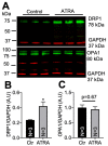
DRP1 and OPA1 levels in mouse hearts treated with an i.p. injection of ATRA or corn oil (vehicle control). (A) Western blots of DRP1 (upper panel) and OPA1 (lower panel). (B) DRP1 levels were significantly higher in ATRA-treated mice (* p < 0.05, vs. vehicle; unpaired 2-sample t-test). (C) There was no effect of ATRA on OPA1.
Figure 3
Figure 3
Quantification of mitochondrial localization of DRP1 and OPA1 by confocal microscopy. (A) Mitochondria were labeled with antibodies against DRP1 and (C) OPA1 in purified cardiac mitochondrial clusters from vehicle- (upper panels) and ATRA- (lower panels) treated mice. Red channel: MitoTracker™ Red. (B) and (D) Mander’s coefficient of colocalization between MitoTracker™ Red (M2; black bars) and DRP1 (M1; gray bars) vs. OPA1 (M1; gray bars), respectively. In ATRA treatment, there was a significant increase in DRP1 colocalization with MitoTracker™ Red (* p < 0.01, vs. vehicle; unpaired 2-sample t-test; n = 3 mice; n = 12 images) (B) but no change in OPA1 (n = 3 mice; n = 12 images) (D). Mitochondria were loaded with MitoTracker™ Red (E) and labeled with secondary antibody alone (F).
Figure 4
Figure 4
Effects of ATRA on mitochondrial ultrastructure in mice treated with ATRA versus control. (A) Representative electron micrographs from ATRA- and corn oil- (vehicle) treated mice at 10,000×, 25,000×, and 50,000× magnification. Mitochondrial (B), average area (C), perimeter and (D) circularity, and (E) number of mitochondria per area. Mitochondrial area and perimeter were significantly decreased (B,C) and mitochondria were more circular in the ATRA-treated group (D) (* p < 0.05, *** p < 0.001, vs. vehicle; unpaired 2-sample t-test). n = 3 hearts; n = 9 micrographs each condition.
Figure 5
Figure 5
Microscopic properties of the mitochondrial network in control and ATRA-treated HL-1 cells. Representative confocal images of the mitochondrial network in (A) control and (B) ATRA-treated cells from experiments (left panels) and the network retrieved using image processing (right panels). Distributions of loop sizes (C), branch lengths (D), and cluster sizes (E) (cumulative probability) for control (red) and ATRA-treated cells (blue). Mean degree (F), average cluster size (G), giant cluster (H), and the giant cluster normalized to the total network size (I). * p < 0.05, ** p < 0.01, vs. control; unpaired 2-sample t-test.
Figure 6
Figure 6
Estimating relative fusion and fission rates using agent-based model. (A) Model scheme representing the tip-to-tip fusion of two X1 nodes into X2. Tip-to-side fusion of one X1 node with one X2 node to form one X3 node, as well as the corresponding fission processes. (B) The number of vehicle (control), or ATRA X1, X2, and X3 species from the model as functions of the number of iterations using the C1 and C2 values given in Table 1. Model results for mean degree (C) and Ng/N (D) as functions of C2 at fixed C1 = 0.0007 and n = 3000. (E) Comparison of Ng/N versus <k> values obtained from experiment (red bullets) and simulation (black crosses) in control and ATRA-treated cells.

Similar articles

Cited by

References

    1. Osellame L.D., Blacker T.S., Duchen M.R. Cellular and molecular mechanisms of mitochondrial function. Best Pract. Res. Clin. Endocrinol. Metab. 2012;26:711–723. doi: 10.1016/j.beem.2012.05.003. - DOI - PMC - PubMed
    1. Carreira R.S., Billups P., Gottlieb R.A. Mitochondrial therapeutics for cardioprotection. Curr. Pharm. Des. 2011;17:2017–2035. doi: 10.2174/138161211796904777. - DOI - PMC - PubMed
    1. Chen H., Chen H., Detmer S.A., Ewald A.J., Griffin E.E., Fraser S.E., Chan D.C. Mitofusins Mfn1 and Mfn2 coordinately regulate mitochondrial fusion and are essential for embryonic devel-opment. J. Cell Biol. 2003;160:189–200. doi: 10.1083/jcb.200211046. - DOI - PMC - PubMed
    1. Smirnova E., Griparic L., Shurland D.-L., Van Der Bliek A.M. Dynamin-related Protein Drp1 Is Required for Mitochondrial Division in Mammalian Cells. Mol. Biol. Cell. 2001;12:2245–2256. doi: 10.1091/mbc.12.8.2245. - DOI - PMC - PubMed
    1. Labrousse A.M., Zappaterra M.D., Rube D.A., van der Bliek A.M. C. elegans Dynamin-Related Protein DRP-1 Controls Severing of the Mitochondrial Outer Membrane. Mol. Cell. 1999;4:815–826. doi: 10.1016/S1097-2765(00)80391-3. - DOI - PubMed

Publication types

LinkOut - more resources