. 2021 May 14;22(10):5201.
doi: 10.3390/ijms22105201.

High Flexibility of RNaseH2 Catalytic Activity with Respect to Non-Canonical DNA Structures

Affiliations

High Flexibility of RNaseH2 Catalytic Activity with Respect to Non-Canonical DNA Structures

Maria Dede et al. Int J Mol Sci. .

Abstract

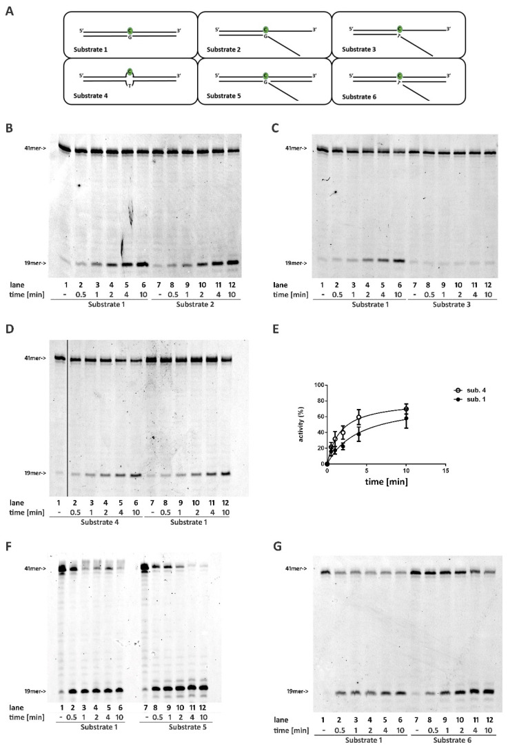
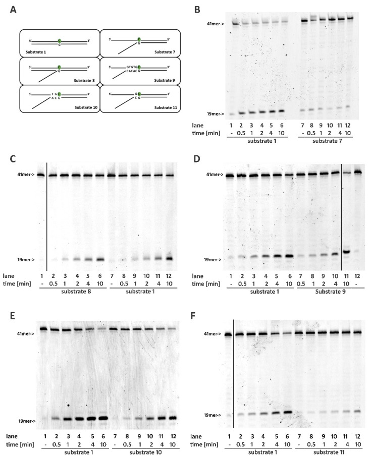
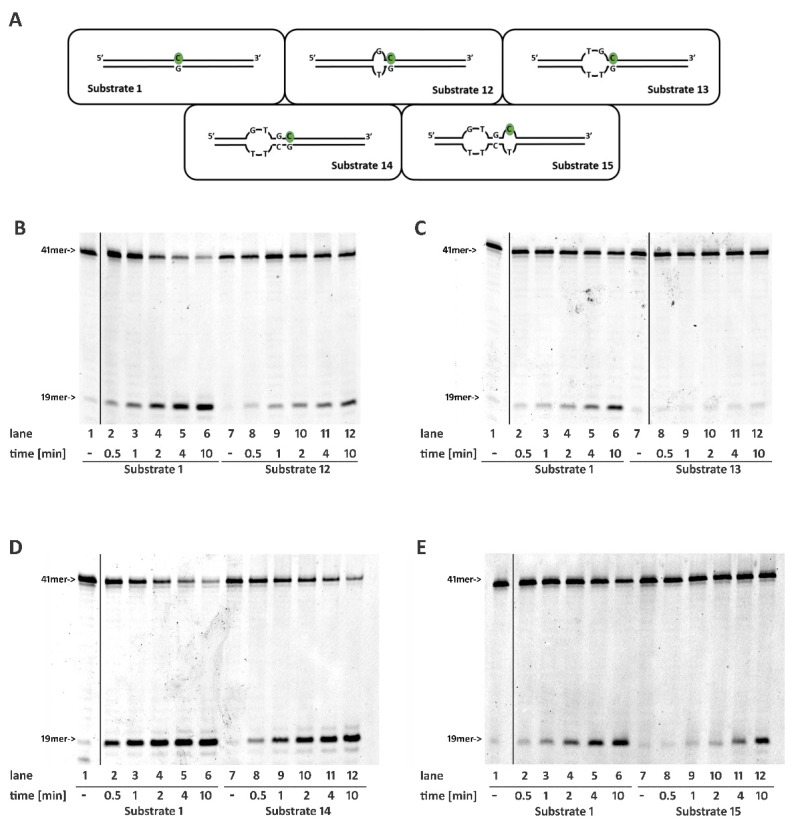
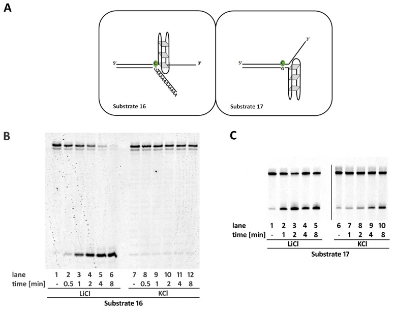
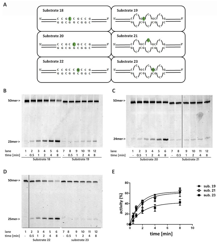
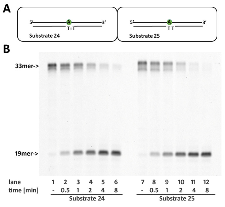
Ribonucleotides misincorporated in the human genome are the most abundant DNA lesions. The 2'-hydroxyl group makes them prone to spontaneous hydrolysis, potentially resulting in strand breaks. Moreover, their presence may decrease the rate of DNA replication causing replicative fork stalling and collapse. Ribonucleotide removal is initiated by Ribonuclease H2 (RNase H2), the key player in Ribonucleotide Excision Repair (RER). Its absence leads to embryonic lethality in mice, while mutations decreasing its activity cause Aicardi-Goutières syndrome. DNA geometry can be altered by DNA lesions or by peculiar sequences forming secondary structures, like G-quadruplex (G4) and trinucleotide repeats (TNR) hairpins, which significantly differ from canonical B-form. Ribonucleotides pairing to lesioned nucleotides, or incorporated within non-B DNA structures could avoid RNase H2 recognition, potentially contributing to genome instability. In this work, we investigate the ability of RNase H2 to process misincorporated ribonucleotides in a panel of DNA substrates showing different geometrical features. RNase H2 proved to be a flexible enzyme, recognizing as a substrate the majority of the constructs we generated. However, some geometrical features and non-canonical DNA structures severely impaired its activity, suggesting a relevant role of misincorporated ribonucleotides in the physiological instability of specific DNA sequences.

Keywords: RER; RNaseH2; misincorporated ribonucleotides; non-B DNA.


Conflict of interest statement

The authors declare no conflict of interest.

Figures

Figure 1
Figure 1
Base pairing at the 3′ of an embedded ribonucleotide is not required for RNaseH2 activity. Time course reactions were performed as described in materials and methods using 20 nM nucleic acid substrates and 0,4 nM RNase H2. (A) Schematic representation of substrates used. Enzyme activity was compared between (B) Substrates 1 and 2; (C) Substrates 1 and 3; (D) Substrates 1 and 4; (F) Substrates 1 and 5; (G) Substrates 1 and 6. Unprocessed substrates and reaction products are indicated at the left of each panel (41-mer and 19-mer respectively). Black vertical line in (D) represents the cropped area in the raw picture. (E) Quantification of three independent experiments measuring RNase H2 activity using substrate 1 and 4.
Figure 2
Figure 2
Base pairing at the 5′ of an embedded ribonucleotide is essential for RNase H2 activity. Time course reactions were performed as described in materials and methods using 20 nM nucleic acid substrates and 0.4 nM RNase H2. (A) Schematic representation of substrates used. Enzyme activity was compared between (B) Substrates 1 and 7; (C) Substrates 1 and 8; (D) Substrates 1 and 9; (E) Substrates 1 and 10; (F) Substrates 1 and 11. Unprocessed substrates and reaction products are indicated at the left of each panel (41-mer and 19-mer respectively). Black vertical lines represent cropped areas in the raw pictures.
Figure 3
Figure 3
Correct pairing of the two nucleotides flanking the 5′ of the embedded ribonucleotide is essential for RNase H2 activity. Time course reactions were performed as described in materials and methods using 20 nM nucleic acid substrates and 0.4 nM RNaseH2. (A) Schematic representation of substrates used. Enzyme activity was compared between (B) Substrates 1 and 12; (C) Substrates 1 and 13; (D) Substrates 1 and 14; (E) Substrates 1 and 15. Unprocessed substrates and reaction products are indicated at the left of each panel (41mer and 19mer respectively). Black vertical lines represent cropped areas in the raw pictures.
Figure 4
Figure 4
A G4 structure on the same strand and flanking the 3′ of a single ribonucleotide impairs RNase H2 activity. (A) schematic representation of Substrates 16 and 17 annealed in the presence of KCl. Time course reactions were performed as described in materials and methods using 20 nM of Substrate 16 (B), or 17 (C), annealed in the presence of KCl (allowing G4 formation), or in the presence of LiCl (not allowing G4 formation). Unprocessed substrates and reaction products are indicated at the left of each panel (44mer and 22mer respectively).
Figure 5
Figure 5
Ribonucleotides embedded in structures mimicking the stem part of CCG hairpins are poor substrates of RNase H2. (A) Schematic representation of substrates mimicking the stem of CCG hairpin structures. Time course reactions were performed using 20 nM nucleic acid substrates and 0.4 nM RNase H2. Enzyme activity was compared between (B) Substrates 18 and 19; (C) Substrates 20 and 21; (D) Substrates 22 and 23. (E) Quantification of three independent RNase H2 reactions using Substrates 19, 21 and 23. Unprocessed substrates and reaction products are indicated at the left of each panel (50mer and 25mer respectively). Black vertical lines in (C,D) represents cropped areas in the raw pictures.
Figure 6
Figure 6
The presence of a CPD lesion opposite a single ribonucleotide does not affects RNase H2 activity. (A) Schematic representation of Substrates 24 (CTD) and 25 (TT). (B) Enzyme activity was compared between Substrates 24 and 25. Time course reactions were performed as described in materials and methods using 0.4 nM RNase H2 and 20 nM Substrates 24 and 25 as indicated.

Similar articles

Cited by

References

    1. Caldecott K.W. Molecular biology. Ribose--An internal threat to DNA. Science. 2014;343:260–261. doi: 10.1126/science.1248234. - DOI - PubMed
    1. Nick McElhinny S.A., Watts B.E., Kumar D., Watt D.L., Lundström E.B., Burgers P.M., Johansson E., Chabes A., Kunkel T.A. Abundant ribonucleotide incorporation into DNA by yeast replicative polymerases. Proc. Natl. Acad. Sci. USA. 2010;107:4949–4954. doi: 10.1073/pnas.0914857107. - DOI - PMC - PubMed
    1. Yao N.Y., Schroeder J.W., Yurieva O., Simmons L.A., O’Donnell M.E. Cost of rNTP/dNTP pool imbalance at the replication fork. Proc. Natl. Acad. Sci. USA. 2013;110:12942–12947. doi: 10.1073/pnas.1309506110. - DOI - PMC - PubMed
    1. Ghodgaonkar M.M., Lazzaro F., Olivera-Pimentel M., Artola-Borán M., Cejka P., Reijns M.A., Jackson A.P., Plevani P., Muzi-Falconi M., Jiricny J. Ribonucleotides misincorporated into DNA act as strand-discrimination signals in eukaryotic mismatch repair. Mol. Cell. 2013;50:323–332. doi: 10.1016/j.molcel.2013.03.019. - DOI - PMC - PubMed
    1. Lujan S.A., Williams J.S., Clausen A.R., Clark A.B., Kunkel T.A. Ribonucleotides are signals for mismatch repair of leading-strand replication errors. Mol. Cell. 2013;50:437–443. doi: 10.1016/j.molcel.2013.03.017. - DOI - PMC - PubMed

LinkOut - more resources