Review
. 2021 May 14;22(10):5200.
doi: 10.3390/ijms22105200.

Structural Plasticity of LL-37 Indicates Elaborate Functional Adaptation Mechanisms to Bacterial Target Structures

Affiliations
Review

Structural Plasticity of LL-37 Indicates Elaborate Functional Adaptation Mechanisms to Bacterial Target Structures

Kornelius Zeth et al. Int J Mol Sci. .

Abstract

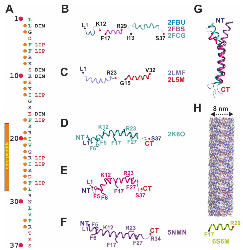
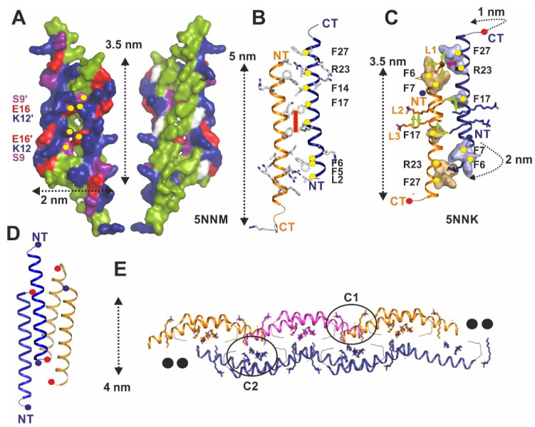
The human cathelicidin LL-37 is a multifunctional peptide of the human innate immune system. Among the various functions of LL-37, its antimicrobial activity is important in controlling the microorganisms of the human body. The target molecules of LL-37 in bacteria include membrane lipids, lipopolysaccharides (LPS), lipoteichoic acid (LTA), proteins, DNA and RNA. In this mini-review, we summarize the entity of LL-37 structural data determined over the last 15 years and specifically discuss features implicated in the interactions with lipid-like molecules. For this purpose, we discuss partial and full-length structures of LL-37 determined in the presence of membrane-mimicking detergents. This constantly growing structural database is now composed of monomers, dimers, tetramers, and fiber-like structures. The diversity of these structures underlines an unexpected plasticity and highlights the conformational and oligomeric adaptability of LL-37 necessary to target different molecular scaffolds. The recent co-crystal structures of LL-37 in complex with detergents are particularly useful to understand how these molecules mimic lipids and LPS to induce oligomerization and fibrillation. Defined detergent binding sites provide deep insights into a new class of peptide scaffolds, widening our view on the fascinating world of the LL-37 structural factotum. Together, the new structures in their evolutionary context allow for the assignment of functionally conserved residues in oligomerization and target interactions. Conserved phenylalanine and arginine residues primarily mediate those interactions with lipids and LPS. The interactions with macromolecules such as proteins or DNA remain largely unexplored and open a field for future studies aimed at structures of LL-37 complexes. These complexes will then allow for the structure-based rational design of LL-37-derived peptides with improved antibiotic properties.

Keywords: LL-37; antimicrobial peptides; cathelicidin; structural flexibility.


Conflict of interest statement

The authors declare no conflict of interest.

Figures

Figure 1
Figure 1
Monomeric full-length and truncated structures of LL-37 determined in the presence of detergents are all α-helical. (A) Sequence of LL-37 to visualize hydrophobic residues color-coded in green, positively charged residues in blue, negatively charged residues in red, and hydrophilic uncharged residues in magenta. Combined evolutionary conservation and structure-based functional assignment are given. Orange dots on the left side of the sequence (marked by numbers 1–37) indicate high sequence identity, and structure-based functions for residues are assigned on the right (DIM—dimerization interface; LIP—lipid-binding site). (B) Structures of LL-12 (Leu1-Lys12), FK-13 (Phe17-Arg29), and IG-25 (Ile13-Ser37) determined in the presence of sodium dodecyl sulfate (SDS) or dioctanoyl phosphatidylglycerol (D8PG) by nuclear magnetic resonance (NMR) in cartoon representation and color-coded in three different colors with N-terminus (NT), and C-terminus (CT) are marked. The first and last residue is given together with the PDB-entry code. (C) Subsequently, published structures of LL-37 fragments solved by NMR, including the antimicrobial inactive LL-23 (Leu1-Arg23) mutant derivative, show α-helical structures. (D) The full-length structure of LL-37 determined in the presence of SDS by NMR shows a bent two-partite structure (residues implicated in detergent binding are shown in stick representation). (E) Full-length structure of LL-37 in dodecylphosphocholine (DPC) determined by NMR displays defined detergent-peptide interactions and a kinked α-helix. (F) Crystal structure of LL-37 determined in the presence of DPC shows defined detergent binding sites representing lipid and lipopolysaccharide (LPS) binding sites in vivo, respectively. The structure shows a disordered but structured part at the N-terminus (residues Leu1-Arg7). (G) Superposition of the three LL-37 structures (see (DF)) shows a significant structural deviation at the N-terminus. (H) Structure of the LL-37 (Phe17-Arg29) derivative crystallized in a packing that allowed the construction of fibers.
Figure 2
Figure 2
Crystal structures of the dimeric and tetrameric full-length LL-37 show significant structural rearrangement. (A) Surface structure representation of LL-37 crystallized in the absence of detergents (PDB-entry: 5NNM). There are two faces of the peptide, one of which is strongly charged (surplus +12/dimer; Arg and Lys are in blue, Asp and Glu in red and Tyr, Ser, Thr, Asn, Gln in magenta), while the opposite side is hydrophobic (all hydrophobic residues are color-coded green). Hydrophilic residues at the interface forming H-bonds are marked by yellow dots. (B) Structure of LL-37 is determined in the absence of detergents. The 5 nm long peptide complex is shown in the same orientation as (A) with the N- (NT), the C-termini (CT), and the twofold symmetry axis (red square) marked. Conserved residues at the dimer interface are marked with yellow dots, according to the published analysis [22]. (C) Structure of LL-37 is determined in the presence of LDAO. Three detergent molecules (L1-L3) per monomer could be identified in the structure, two of which in the center of the dimer with the alkyl chains oriented perpendicular to the peptide chains and interactions between detergent and LL-37 are marked by green arrows. One detergent was observed between the head-to-tail arranged dimers in the crystal packing (see also (E)). (D) Tetrameric channel structure of LL-37 formed by two antiparallel dimers (shown in orange and blue). (E) Head-to-tail polymerization and fiber formation (diameter 4 nm) of LL-37 crystallized in the presence of LDAO and DPC. Two LDAO detergent molecules are encapsulated in the pocket, which is formed after polymerization of the dimeric peptide. There are two reoccurring clusters of detergents observed along the fibril, one between the head and tails of the polymer (C1) and another one at the center of the dimers (C2).

Similar articles

Cited by

References

    1. Alanis A.J. Resistance to Antibiotics: Are We in the Post-Antibiotic Era? Arch. Med. Res. 2005;36:697–705. doi: 10.1016/j.arcmed.2005.06.009. - DOI - PubMed
    1. Zasloff M. Antimicrobial peptides of multicellular organisms. Nat. Cell Biol. 2002;415:389–395. doi: 10.1038/415389a. - DOI - PubMed
    1. Koo H.B., Seo J. Antimicrobial peptides under clinical investigation. Pept. Sci. 2019;111:e24122. doi: 10.1002/pep2.24122. - DOI
    1. Peschel A., Sahl H.-G. The co-evolution of host cationic antimicrobial peptides and microbial resistance. Nat. Rev. Genet. 2006;4:529–536. doi: 10.1038/nrmicro1441. - DOI - PubMed
    1. Ventola C.L. The antibiotic resistance crisis: Part 2: Management strategies and new agents. Pharm. Ther. 2015;40:344–352. - PMC - PubMed

LinkOut - more resources