. 2021 May 14;9(5):508.
doi: 10.3390/vaccines9050508.

M448R and MGF505-7R: Two African Swine Fever Virus Antigens Commonly Recognized by ASFV-Specific T-Cells and with Protective Potential

Affiliations

M448R and MGF505-7R: Two African Swine Fever Virus Antigens Commonly Recognized by ASFV-Specific T-Cells and with Protective Potential

Laia Bosch-Camós et al. Vaccines (Basel). .

Abstract

African swine fever (ASF) is today's number one threat for the global swine industry. Neither commercial vaccine nor treatment is available against ASF and, thus far, only live attenuated viruses (LAV) have provided robust protection against lethal ASF virus (ASFV) challenge infections. Identification of ASFV proteins inducing protective immune responses is one of the major challenges to develop safer and efficient subunit vaccines. Immunopeptidomic studies recently performed in our laboratory allowed identifying ASFV antigens recognized by ASFV-specific CD8+ T-cells. Here, we used data from the SLAI-peptide repertoire presented by a single set of ASFV-infected porcine alveolar macrophages to generate a complex DNA vaccine composed by 15 plasmids encoding the individual peptide-bearing ORFs. DNA vaccine priming improved the protection afforded by a suboptimal dose of the BA71ΔCD2 LAV given as booster vaccination, against Georgia2007/1 lethal challenge. Interestingly, M448R was the only protein promiscuously recognized by the induced ASFV-specific T-cells. Furthermore, priming pigs with DNA plasmids encoding M488R and MGF505-7R, a CD8+ T-cell antigen previously described, confirmed these two proteins as T-cell antigens with protective potential. These studies might be useful to pave the road for designing safe and more efficient vaccine formulations in the future.

Keywords: African swine fever; DNA immunization; T-cells; antigen discovery; immunopeptidomics; live attenuated virus; protection.


Conflict of interest statement

The authors declare no conflict of interest.

Figures

Figure 1
Figure 1
Schematic representation of the in vivo experimental designs. The two in vivo experiments performed in this study followed an identical scheme but priming with different plasmid combinations: either 15 clones in Experiment 1 or the combination of two plasmids (pCMV-Ub-M448R + pCMV-Ub-MGF505-7R) in Experiment 2. Groups of five pigs were primed twice two weeks apart using the indicated DNA plasmid mixes and boosted with 103 PFU of BA71∆CD2. Three weeks after the boost, pigs were challenged with a lethal dose of Georgia2007/1. Samples were taken at different days post priming (dpp), post boost (dpb) or post challenge (dpc).
Figure 2
Figure 2
Priming pigs with 15 plasmids improves the protection induced by suboptimal BA71ΔCD2 immunization against lethal Georgia2007/1 challenge. Pigs were immunized twice with either the empty pCMV-Ub plasmid (Control) or the 15 plasmids and next boosted with 103 PFU BA71ΔCD2. Two weeks later, all pigs were challenged with a lethal dose of Georgia2007/1 and (A) deaths, (B) ASF-compatible clinical signs and (C) rectal temperature from 15 plasmids (top) and control (bottom) groups were recorded daily. Solid lines represent animals that survived the challenge while dashed lines symbolize animals that succumbed the challenge.
Figure 3
Figure 3
ASFV virus titers in sera and nasal swabs found in pigs after Georgia2007/1 challenge. (A) ASFV titers as indicated by GEC/mL found in sera and (B) nasal swabs analyzed by qPCR at different time points post challenge in the 15 plasmids group (top) and the control group (bottom). Solid lines represent animals that survived the challenge while dashed lines indicate animals that succumbed to the challenge.
Figure 4
Figure 4
M488R is frequently recognized by ASFV-specific T-cells. (A) ASFV-specific antibodies (total IgG) were measured by ELISA, expressing the results as OD values at a wavelength of 450 nm. Solid lines represent the 15-ASFV plasmids group and dashed lines represent the control group. (B) ASFV-specific T-cell responses were assessed by IFNΥ ELISpot using PBMCs isolated at different time points: after DNA prime (14 dpp), after BA71∆CD2 boost (21 dpb) and after Georgia2007/1 challenge (21 dpc). (C) IFNΥ ELISpot using PBMCs from surviving animals as effector cells and autologous fibroblasts transfected with pCMV-Ub-M448R or the 15 plasmids as specific stimuli. indicates animals succumbing to ASFV challenge.
Figure 5
Figure 5
Priming pigs with pCMV-Ub-M448R and pCMV-Ub-MGF505-7R improves the protection afforded by suboptimal BA71ΔCD2 immunization against lethal Georgia2007/1 challenge. Pigs were immunized twice with either the empty pCMV-Ub plasmid (Control) or pCMV-Ub-M488R + pCMV-Ub-MGF505-7R and next boosted with 103 PFU BA71ΔCD2. Two weeks later, all pigs were challenged with a lethal dose of Georgia2007/1 and (A) deaths, (B) ASF typical clinical signs and (C) rectal temperature from pCMV-Ub-M488R + pCMV-Ub-MGF505-7R (top) and control (bottom) groups were recorded daily. Solid lines represent animals that survived the challenge while dashed lines symbolize animals that succumbed to the challenge.
Figure 6
Figure 6
Virus DNA titers found in sera and nasal swabs of pigs after Georgia2007/1 challenge. (A) ASFV GEC titers found in sera and (B) nasal swabs at different times post Georgia2007/1 challenge detected by qPCR in the pCMV-Ub-M448R and pCMV-Ub-MGF505-7R primed group (top) and the control group (bottom). Solid lines represent animals that survived the challenge while dashed lines symbolize animals that succumbed to the challenge.
Figure 7
Figure 7
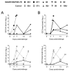
M488R and MGF505-7R are frequently recognized by specific T-cells. (A) ASFV-specific antibodies (total IgG) were measured by ELISA, expressing the results as OD values at a wavelength of 450 nm. Solid lines represent the M448R + MGF505-7R group and dashed lines represent the control group. ASFV-specific T-cell responses were assessed by IFNΥ ELISpot using as stimulus (B) ASFV or (C) autologous fibroblasts transfected with either pCMV-Ub-M448R or pCMV-Ub-MGF505-7R at different time points: after DNA prime (14 dpp), after BA71∆CD2 boost (21 dpb) and after Georgia2007/1 challenge (21 dpc). Data from the M448R+MGF505-7R primed group and control group are represented at the top and bottom, respectively. indicates animals succumbing to ASFV challenge.

References

    1. Beltran-Alcrudo D., Lubroth J., Depner K., La Rocque S. African swine fever in the Caucasus. FAO Empres. Watch. 2008;1:1–8.
    1. Cwynar P., Stojkov J., Wlazlak K. African Swine Fever Status in Europe. Viruses. 2019;11:310. doi: 10.3390/v11040310. - DOI - PMC - PubMed
    1. Gogin A., Gerasimov V., Malogolovkin A., Kolbasov D. African swine fever in the North Caucasus region and the Russian Federation in years 2007–2012. Virus Res. 2013;173:198–203. doi: 10.1016/j.virusres.2012.12.007. - DOI - PubMed
    1. Kim S.H., Kim J., Son K., Choi Y., Jeong H.S., Kim Y.K., Park J.E., Hong Y.J., Lee S.I., Wang S.J., et al. Wild boar harbouring African swine fever virus in the demilitarized zone in South Korea, 2019. Emerg. Microbes Infect. 2020;9:628–630. doi: 10.1080/22221751.2020.1738904. - DOI - PMC - PubMed
    1. Li X., Tian K. African swine fever in China. Vet. Rec. 2018;183:300–301. doi: 10.1136/vr.k3774. - DOI - PubMed