

Structural Insights into the Interaction of Filovirus Glycoproteins with the Endosomal Receptor Niemann-Pick C1: A Computational Study
- PMID: 34069246
- PMCID: PMC8156010
- DOI: 10.3390/v13050913
Structural Insights into the Interaction of Filovirus Glycoproteins with the Endosomal Receptor Niemann-Pick C1: A Computational Study
Abstract
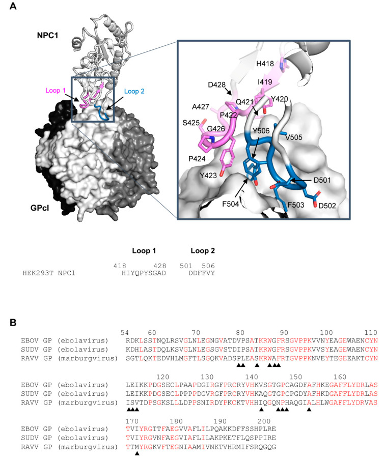
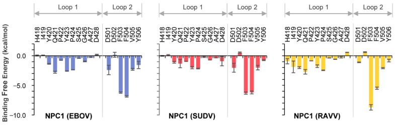
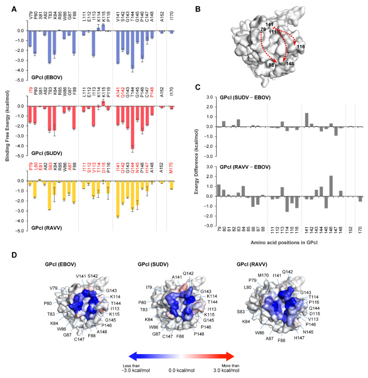
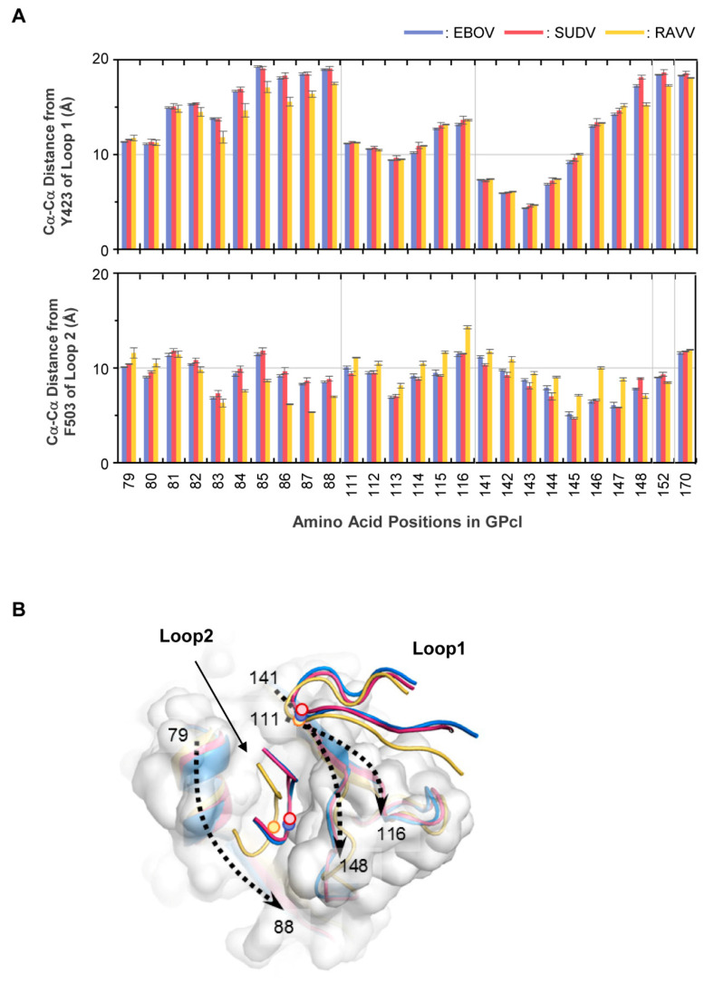
Filoviruses, including marburgviruses and ebolaviruses, have a single transmembrane glycoprotein (GP) that facilitates their entry into cells. During entry, GP needs to be cleaved by host proteases to expose the receptor-binding site that binds to the endosomal receptor Niemann-Pick C1 (NPC1) protein. The crystal structure analysis of the cleaved GP (GPcl) of Ebola virus (EBOV) in complex with human NPC1 has demonstrated that NPC1 has two protruding loops (loops 1 and 2), which engage a hydrophobic pocket on the head of EBOV GPcl. However, the molecular interactions between NPC1 and the GPcl of other filoviruses remain unexplored. In the present study, we performed molecular modeling and molecular dynamics simulations of NPC1 complexed with GPcls of two ebolaviruses, EBOV and Sudan virus (SUDV), and one marburgvirus, Ravn virus (RAVV). Similar binding structures were observed in the GPcl-NPC1 complexes of EBOV and SUDV, which differed from that of RAVV. Specifically, in the RAVV GPcl-NPC1 complex, the tip of loop 2 was closer to the pocket edge comprising residues at positions 79-88 of GPcl; the root of loop 1 was predicted to interact with P116 and Q144 of GPcl. Furthermore, in the SUDV GPcl-NPC1 complex, the tip of loop 2 was slightly closer to the residue at position 141 than those in the EBOV and RAVV GPcl-NPC1 complexes. These structural differences may affect the size and/or shape of the receptor-binding pocket of GPcl. Our structural models could provide useful information for improving our understanding the differences in host preference among filoviruses as well as contributing to structure-based drug design.
Keywords: Niemann-Pick C1; ebolavirus; filovirus; glycoprotein; marburgvirus; molecular dynamics; molecular modeling; structure.
Conflict of interest statement
The authors declare no conflict of interest.
Figures
Similar articles
-
Direct Intracellular Visualization of Ebola Virus-Receptor Interaction by In Situ Proximity Ligation.mBio. 2021 Jan 12;12(1):e03100-20. doi: 10.1128/mBio.03100-20. mBio. 2021. PMID: 33436438 Free PMC article.
-
Ebola Viral Glycoprotein Bound to Its Endosomal Receptor Niemann-Pick C1.Cell. 2016 Jan 14;164(1-2):258-268. doi: 10.1016/j.cell.2015.12.044. Cell. 2016. PMID: 26771495 Free PMC article.
-
Filoviruses Use the HOPS Complex and UVRAG To Traffic to Niemann-Pick C1 Compartments during Viral Entry.J Virol. 2020 Jul 30;94(16):e01002-20. doi: 10.1128/JVI.01002-20. Print 2020 Jul 30. J Virol. 2020. PMID: 32493822 Free PMC article.
-
Potential pharmacological strategies targeting the Niemann-Pick C1 receptor and Ebola virus glycoprotein interaction.Eur J Med Chem. 2021 Nov 5;223:113654. doi: 10.1016/j.ejmech.2021.113654. Epub 2021 Jun 19. Eur J Med Chem. 2021. PMID: 34175537 Review.
-
Filovirus entry: a novelty in the viral fusion world.Viruses. 2012 Feb;4(2):258-75. doi: 10.3390/v4020258. Epub 2012 Feb 7. Viruses. 2012. PMID: 22470835 Free PMC article. Review.
Cited by
-
Navigating the Complex Landscape of Ebola Infection Treatment: A Review of Emerging Pharmacological Approaches.Infect Dis Ther. 2024 Jan;13(1):21-55. doi: 10.1007/s40121-023-00913-y. Epub 2024 Jan 19. Infect Dis Ther. 2024. PMID: 38240994 Free PMC article. Review.
-
Unique Mode of Antiviral Action of a Marine Alkaloid against Ebola Virus and SARS-CoV-2.Viruses. 2022 Apr 15;14(4):816. doi: 10.3390/v14040816. Viruses. 2022. PMID: 35458549 Free PMC article.
-
Molecular determinants of cross-species transmission in emerging viral infections.Microbiol Mol Biol Rev. 2024 Sep 26;88(3):e0000123. doi: 10.1128/mmbr.00001-23. Epub 2024 Jun 24. Microbiol Mol Biol Rev. 2024. PMID: 38912755 Free PMC article. Review.
-
Decoding the blueprint of receptor binding by filoviruses through large-scale binding assays and machine learning.Cell Host Microbe. 2025 Feb 12;33(2):294-313.e11. doi: 10.1016/j.chom.2024.12.016. Epub 2025 Jan 15. Cell Host Microbe. 2025. PMID: 39818205
References
-
- Kuhn J.H., Adkins S., Alioto D., Alkhovsky S.V., Amarasinghe G.K., Anthony S.J., Avšič-Županc T., Ayllón M.A., Bahl J., Balkema-Buschmann A., et al. 2020 taxonomic update for phylum Negarnaviricota (Riboviria: Orthornavirae), including the large orders Bunyavirales and Mononegavirales. Arch. Virol. 2020;165:3023–3072. doi: 10.1007/s00705-020-04731-2. - DOI - PMC - PubMed
-
- Sebastian S., Flaxman A., Cha K.M., Ulaszewska M., Gilbride C., Sharpe H., Wright E., Spencer A.J., Dowall S., Hewson R., et al. A Multi-Filovirus Vaccine Candidate: Co-Expression of Ebola, Sudan, and Marburg Antigens in a Single Vector. Vaccines. 2020;8:241. doi: 10.3390/vaccines8020241. - DOI - PMC - PubMed
-
- Wec A.Z., Herbert A.S., Murin C.D., Nyakatura E.K., Abelson D.M., Fels J.M., He S., James R.M., de La Vega M.-A., Zhu W., et al. Antibodies from a Human Survivor Define Sites of Vulnerability for Broad Protection against Ebolaviruses. Cell. 2017;169:878–890. doi: 10.1016/j.cell.2017.04.037. - DOI - PMC - PubMed
Publication types
MeSH terms
Substances
LinkOut - more resources
Full Text Sources
