. 2021 May 14;10(5):603.
doi: 10.3390/pathogens10050603.

Altered Nasal Microbiota Composition Associated with Development of Polyserositis by Mycoplasma hyorhinis

Affiliations

Altered Nasal Microbiota Composition Associated with Development of Polyserositis by Mycoplasma hyorhinis

Miguel Blanco-Fuertes et al. Pathogens. .

Abstract

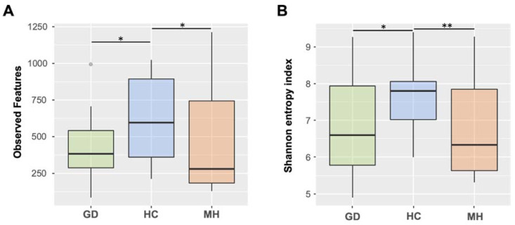
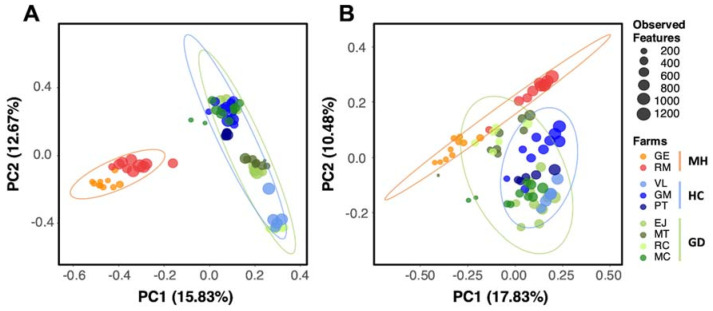
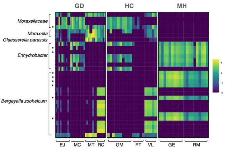
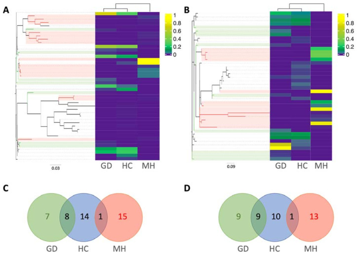
Fibrinous polyserositis in swine farming is a common pathological finding in nursery animals. The differential diagnosis of this finding should include Glaesserella parasuis (aetiological agent of Glässer's disease) and Mycoplasma hyorhinis, among others. These microorganisms are early colonizers of the upper respiratory tract of piglets. The composition of the nasal microbiota at weaning was shown to constitute a predisposing factor for the development of Glässer's disease. Here, we unravel the role of the nasal microbiota in the subsequent systemic infection by M. hyorhinis, and the similarities and differences with Glässer's disease. Nasal samples from farms with recurrent problems with polyserositis associated with M. hyorhinis (MH) or Glässer's disease (GD) were included in this study, together with healthy control farms (HC). Nasal swabs were taken from piglets in MH farms at weaning, before the onset of the clinical outbreaks, and were submitted to 16S rRNA gene amplicon sequencing (V3-V4 region). These sequences were analyzed together with sequences from similar samples previously obtained in GD and HC farms. Animals from farms with disease (MH and GD) had a nasal microbiota with lower diversity than those from the HC farms. However, the composition of the nasal microbiota of the piglets from these disease farms was different, suggesting that divergent microbiota imbalances may predispose the animals to the two systemic infections. We also found variants of the pathogens that were associated with the farms with the corresponding disease, highlighting the importance of studying the microbiome at strain-level resolution.

Keywords: 16S rRNA gene; Glässer’s disease; Mycoplasma hyorhinis; microbial diversity; nasal microbiota; porcine polyserositis.


Conflict of interest statement

The authors declare no conflict of interest.

Figures

Figure 1
Figure 1
Boxplots representing median and interquartile ranges of alpha diversity estimated measuring the observed features (A) or Shannon’s index (B) in nasal microbiota from piglets at weaning from healthy control farms (HC) and farms with polyserositis in the nursery caused by Mycoplasma hyorhinis (MH) or caused by Glaesserella parasuis (Glässer’s disease, GD). Outlier is indicated with a grey circle on the plot. Error bars are standard deviation. * means p < 0.05; ** means p ≤ 0.01.
Figure 2
Figure 2
Beta diversity analysis on Bray Curtis (A) and unweighted Unifrac distances (B) of the nasal microbiota of weaning piglets. Each circle represents the microbial composition of each sample and the size of the circles is proportional to the number of Observed Features as indicated in the legend. Healthy control farms (HC) are depicted in blue, while farms with polyserositis caused by M. hyorhinis (MH) are in orange and G. parasuis (GD) in green palettes. Ellipses are calculated with the Euclidean distances of the grouped samples.
Figure 3
Figure 3
Relative abundance in the nasal microbiota of weaning piglets of the top 30 most abundant ASVs among all the groups in log10 scale. The X axis (bottom) shows the distribution of each sample by farm and the Y axis represents the top 30 most abundant ASVs grouped by the taxa assignment. Top labels correspond to the study groups: Glässer’s disease (GD), healthy control (HC) and M. hyorhinis (MH) farms. * Asterisks mark the ASVs detected as statistically different in the differential abundance analysis (ANCOM) among the three study groups (GD, MH, HC).
Figure 4
Figure 4
Relative abundance of M. hyorhinis (A) and G. parasuis (B) amplicon sequence variants (ASVs) in the nasal microbiota of weaning piglets from farms with Glässer’s disease (GD), healthy controls (HC) or farms with polyserositis caused by M. hyorhinis (MH). The phylogenetic relationship of ASVs from each pathogen is represented in Maximum likelihood trees. Branches from ASVs found exclusively in GD farms are colored in green while the ones found only the MH group are colored in red. Venn diagrams representing the distribution of M. hyorhinis (C) and G. parasuis (D) ASVs in the different groups were plotted following the same color pattern.

References

    1. NIH HMP Working Group. Peterson J., Garges S., Giovanni M., McInnes P., Wang L., Schloss J.A., Bonazzi V., McEwen J.E., Wetterstrand K.A., et al. The NIH Human Microbiome Project. Genome Res. 2009;19:2317–2323. doi: 10.1101/gr.096651.109. - DOI - PMC - PubMed
    1. Zheng D., Liwinski T., Elinav E. Interaction between microbiota and immunity in health and disease. Cell Res. 2020;30:492–506. doi: 10.1038/s41422-020-0332-7. - DOI - PMC - PubMed
    1. Han G.G., Lee J.-Y., Jin G.-D., Park J., Choi Y.H., Chae B.J., Kim E.B., Choi Y.-J. Evaluating the association between body weight and the intestinal microbiota of weaned piglets via 16S rRNA Sequencing. Appl. Microbiol. Biotechnol. 2017;101:5903–5911. doi: 10.1007/s00253-017-8304-7. - DOI - PubMed
    1. McCormack U.M., Curião T., Buzoianu S.G., Prieto M.L., Ryan T., Varley P., Crispie F., Magowan E., Metzler-Zebeli B.U., Berry D., et al. Exploring a Possible Link between the Intestinal Microbiota and Feed Efficiency in Pigs. Appl. Environ. Microbiol. 2017;83 doi: 10.1128/AEM.00380-17. - DOI - PMC - PubMed
    1. Man W.H., de Steenhuijsen Piters W.A.A., Bogaert D. The microbiota of the respiratory tract: Gatekeeper to respiratory health. Nat. Rev. Microbiol. 2017;15:259–270. doi: 10.1038/nrmicro.2017.14. - DOI - PMC - PubMed

LinkOut - more resources