Review
. 2021 May 14;13(10):2390.
doi: 10.3390/cancers13102390.

Chondrosarcoma-from Molecular Pathology to Novel Therapies

Affiliations
Review

Chondrosarcoma-from Molecular Pathology to Novel Therapies

Agnieszka E Zając et al. Cancers (Basel). .

Abstract

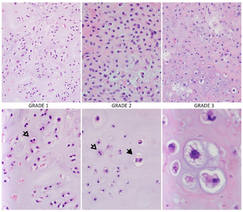
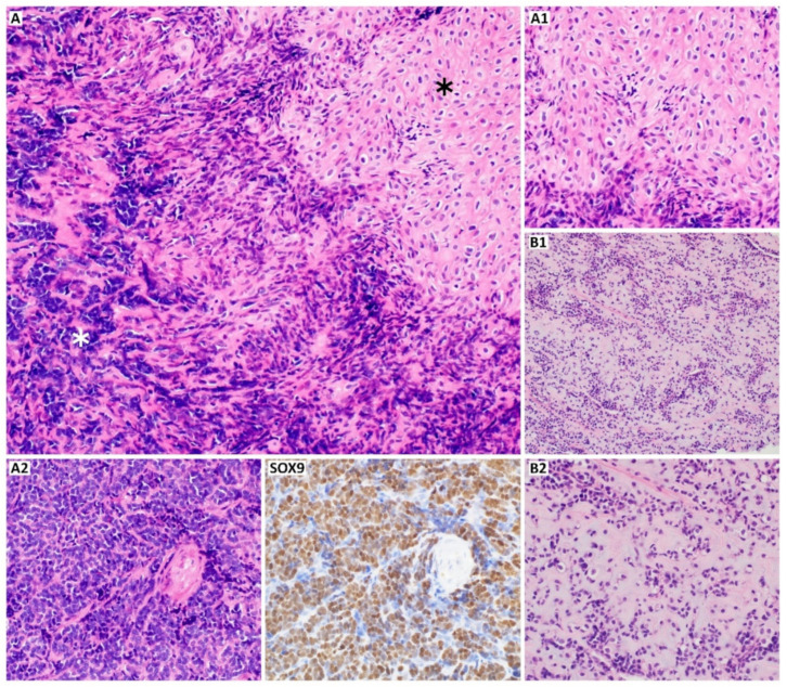
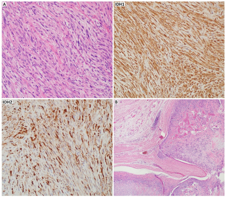
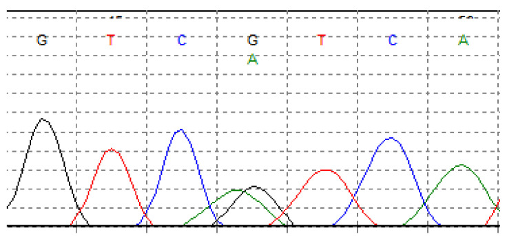
Chondrosarcoma (CHS) is the second most common primary malignant bone sarcoma. Overall survival and prognosis of this tumor are various and often extreme, depending on histological grade and tumor subtype. CHS treatment is difficult, and surgery remains still the gold standard due to the resistance of this tumor to other therapeutic options. Considering the role of differentiation of CHS subtypes and the need to develop new treatment strategies, in this review, we introduced a multidisciplinary characterization of CHS from its pathology to therapies. We described the morphology of each subtype with the role of immunohistochemical markers in diagnostics of CHS. We also summarized the most frequently mutated genes and genome regions with altered pathways involved in the pathology of this tumor. Subsequently, we discussed imaging methods and the role of currently used therapies, including surgery and the limitations of chemo and radiotherapy. Finally, in this review, we presented novel targeted therapies, including those at ongoing clinical trials, which can be a potential future target in designing new therapeutics for patients with CHS.

Keywords: chondrosarcoma; diagnostic markers; pathology; therapies.


Conflict of interest statement

The authors declare no conflict of interest.

Figures

Figure 1
Figure 1
Conventional chondrosarcoma and histological grades. The higher-grade tumors are more hypercellular, with increasing mitotic activity, cytological and nuclear atypia, a matrix is changing from hyaline to more mucoid or myxoid (white arrow: binucleation of chondrocytes, black arrow: large, highly atypical chondrocyte; hematoxylin and eosin staining (HE), magnification: upper row 100×, bottom row 200× and 600×). Photos by Anna Szumera-Ciećkiewicz.
Figure 2
Figure 2
Mesenchymal chondrosarcoma ((A), HE, 200×). A mixture of round cells ((A2), white asterisk,; HE, 200×) with islands of well-differentiated hyaline cartilage ((A1), black asterisk; HE, 200×); the proportions of these two components may vary ((B1), HE, 100×; (B2)), HE, 200×); (SOX9) (SRY-Box Transcription Factor 9, 200×) is highly expressed supporting the differential diagnosis of mesenchymal chondrosarcoma. Photos by Anna Szumera-Ciećkiewicz.
Figure 3
Figure 3
Dedifferentiated and periosteal chondrosarcoma. The transition to undifferentiated pleomorphic sarcoma ((A), HE, 200×), the case was positive with p.Arg132His mutation-specific isocitrate dehydrogenase 1 ((IDH1), 200×) antibody and negative for isocitrate dehydrogenase 2 ((IDH2), 200×); periosteal chondrosarcoma and cortical extension to soft-tissue ((B), HE, 40×). Photos by Anna Szumera-Ciećkiewicz.
Figure 4
Figure 4
Somatic mutation in isocitrate dehydrogenase 1 (IDH1) in chondrosarcoma. G > A transition at nucleotide position 395, in codon 132, leading arginine to histidine substitution (p.Arg132His). Fluorogram by Andrzej Tysarowski.
Figure 5
Figure 5
Somatic mutation in isocitrate dehydrogenase 2 (IDH2) in chondrosarcoma. G > T transition at nucleotide position 516, in codon 172, leading arginine to serine substitution (p.Arg172Ser). Fluorogram by Andrzej Tysarowski.
Figure 6
Figure 6
High-grade chondrosarcoma of the left scapula. Preoperative X-ray of the G3 scapular chondrosarcoma (A) and after en bloc upper interscapulothoracic humeral resection (B) This particular procedure known as Tikhoff-Linberg is a limb-sparing surgical option for tumors in and around the proximal humerus and shoulder girdle and an alternative treatment to forequarter amputation; however, is associated with poor cosmetic appearance (C) that can gradually change over the years depending on soft tissue contracture and scar tissue formation (D). Photos by Bartłomiej Szostakowski.
Figure 7
Figure 7
Low-grade chondrosarcoma. Rarely symptomatic lesions are discovered incidentally on X-rays after injury to the extremity (A,C). Meticulous curettage of G1 chondrosarcomas and polymethyl methacrylate cementoplasty remains the mainstay of treatment (B,D). Photos by Bartłomiej Szostakowski.
Figure 8
Figure 8
High grade locally advanced acral chondrosarcoma. Preoperative image of the locally advanced G2 (A) and G3 (B) chondrosarcoma of the left proximal femur. Reconstruction with modular endoprosthetic replacement (C,D). Photos by Bartłomiej Szostakowski.
Figure 9
Figure 9
Stereotactic body radiotherapy (SBRT) for in-field recurrent high-grade mesenchymal chondrosarcoma (confirmed in next-generation sequencing sarcoma panel). In 2015, the patient underwent neoadjuvant chemotherapy followed by surgery, adjuvant radiochemotherapy (50.4 Gy in 1.8 Gy fractions with vincristine and dactinomycin combination chemotherapy), and chemotherapy according to EIAO regimen (etoposide, ifosfamide, vincristine, and dactinomycin). In 2019, he experienced local recurrence (LR) in the paraspinal area, which was resected in 2020. Five months later, the second LR was observed (A,B). The patients received SBRT (C) planned on fusion with magnetic resonance imaging and previous treatment plan (D). Volumetric arc therapy was used to deliver 30 Gy in five fractions prescribed on 80% isodose (C). Cord tolerance doses were compliant with the report published by Sahgal et al. [162]. Photos by Mateusz J. Spałek.

Similar articles

Cited by

References

    1. Lin P.P., Moussallem C.D., Deavers M.T. Secondary chondrosarcoma. J. Am. Acad. Orthop. Surg. 2010;18:608–615. doi: 10.5435/00124635-201010000-00004. - DOI - PubMed
    1. Valery P.C., Laversanne M., Bray F. Bone cancer incidence by morphological subtype: A global assessment. Cancer Causes Control. 2015;26:1127–1139. doi: 10.1007/s10552-015-0607-3. - DOI - PubMed
    1. Arora R.S., Alston R.D., Eden T.O., Geraci M., Birch J.M. The contrasting age-incidence patterns of bone tumours in teenagers and young adults: Implications for aetiology. Int. J. Cancer. 2012;131:1678–1685. doi: 10.1002/ijc.27402. - DOI - PubMed
    1. Damron T.A., Ward W.G., Stewart A. Osteosarcoma, chondrosarcoma, and Ewing’s sarcoma: National Cancer Data Base Report. Clin. Orthop. Relat. Res. 2007;459:40–47. doi: 10.1097/BLO.0b013e318059b8c9. - DOI - PubMed
    1. Whelan J., McTiernan A., Cooper N., Wong Y.K., Francis M., Vernon S., Strauss S.J. Incidence and survival of malignant bone sarcomas in England 1979–2007. Int. J. Cancer. 2012;131:E508–E517. doi: 10.1002/ijc.26426. - DOI - PubMed

LinkOut - more resources