

The Selective Histone Deacetylase Inhibitor MI192 Enhances the Osteogenic Differentiation Efficacy of Human Dental Pulp Stromal Cells
- PMID: 34069280
- PMCID: PMC8156347
- DOI: 10.3390/ijms22105224
The Selective Histone Deacetylase Inhibitor MI192 Enhances the Osteogenic Differentiation Efficacy of Human Dental Pulp Stromal Cells
Abstract
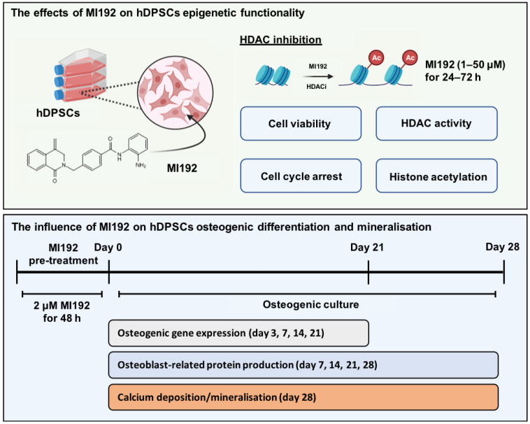
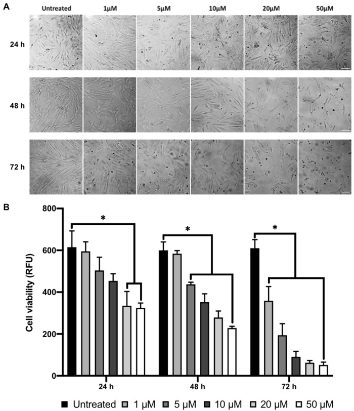
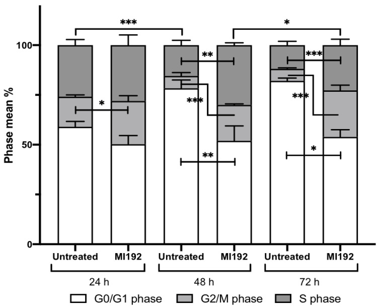
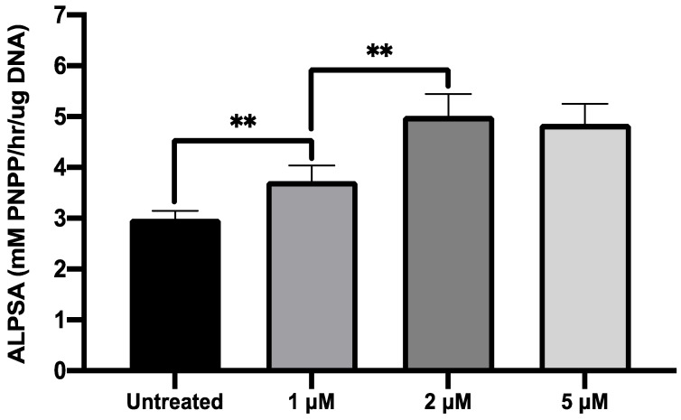
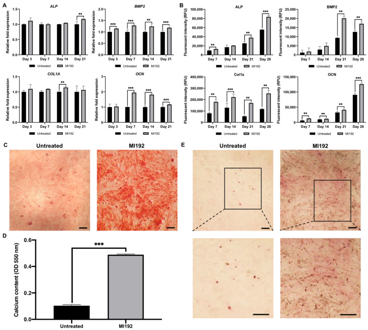
The use of human dental pulp stromal cells (hDPSCs) has gained increasing attention as an alternative stem cell source for bone tissue engineering. The modification of the cells' epigenetics has been found to play an important role in regulating differentiation, with the inhibition of histone deacetylases 3 (HDAC3) being linked to increased osteogenic differentiation. This study aimed to induce epigenetic reprogramming using the HDAC2 and 3 selective inhibitor, MI192 to promote hDPSCs osteogenic capacity for bone regeneration. MI192 treatment caused a time-dose-dependent change in hDPSC morphology and reduction in viability. Additionally, MI192 successfully augmented hDPSC epigenetic functionality, which resulted in increased histone acetylation and cell cycle arrest at the G2/M phase. MI192 pre-treatment exhibited a dose-dependent effect on hDPSCs alkaline phosphatase activity. Quantitative PCR and In-Cell Western further demonstrated that MI192 pre-treatment significantly upregulated hDPSCs osteoblast-related gene and protein expression (alkaline phosphatase, bone morphogenic protein 2, type I collagen and osteocalcin) during osteogenic differentiation. Importantly, MI192 pre-treatment significantly increased hDPSCs extracellular matrix collagen production and mineralisation. As such, for the first time, our findings show that epigenetic reprogramming with the HDAC2 and 3 selective inhibitor MI192 accelerates the osteogenic differentiation of hDPSCs, demonstrating the considerable utility of this MSCs engineering approach for bone augmentation strategies.
Keywords: HDAC inhibitor; MI192; bone tissue engineering; epigenetics; histone deacetylase; human dental pulp stromal cells.
Conflict of interest statement
The authors declare no competing interest.
Figures

Similar articles
-
The effect of epigenetic reprogramming using MI192 HDAC inhibitor on enhancing the osteogenesis of human adipose-derived stem cells in vitro.Biosci Rep. 2023 May 31;43(5):BSR20221635. doi: 10.1042/BSR20221635. Biosci Rep. 2023. PMID: 37022380 Free PMC article.
-
Bone tissue engineering using 3D silk scaffolds and human dental pulp stromal cells epigenetic reprogrammed with the selective histone deacetylase inhibitor MI192.Cell Tissue Res. 2022 Jun;388(3):565-581. doi: 10.1007/s00441-022-03613-0. Epub 2022 Apr 1. Cell Tissue Res. 2022. PMID: 35362831 Free PMC article.
-
MI192 induced epigenetic reprogramming enhances the therapeutic efficacy of human bone marrows stromal cells for bone regeneration.Bone. 2021 Dec;153:116138. doi: 10.1016/j.bone.2021.116138. Epub 2021 Jul 31. Bone. 2021. PMID: 34339909
-
The Role of Histone Acetylation Modification in Dental Tissue-Derived Mesenchymal Stem Cells and Odontogenesis.Cell Reprogram. 2023 Feb;25(1):11-19. doi: 10.1089/cell.2022.0091. Epub 2022 Dec 30. Cell Reprogram. 2023. PMID: 36594932 Review.
-
The Role of Epigenetic in Dental and Oral Regenerative Medicine by Different Types of Dental Stem Cells: A Comprehensive Overview.Stem Cells Int. 2022 Jun 9;2022:5304860. doi: 10.1155/2022/5304860. eCollection 2022. Stem Cells Int. 2022. PMID: 35721599 Free PMC article. Review.
Cited by
-
GelMA Hydrogel Reinforced with 3D Printed PEGT/PBT Scaffolds for Supporting Epigenetically-Activated Human Bone Marrow Stromal Cells for Bone Repair.J Funct Biomater. 2022 Apr 10;13(2):41. doi: 10.3390/jfb13020041. J Funct Biomater. 2022. PMID: 35466223 Free PMC article.
-
The effect of epigenetic reprogramming using MI192 HDAC inhibitor on enhancing the osteogenesis of human adipose-derived stem cells in vitro.Biosci Rep. 2023 May 31;43(5):BSR20221635. doi: 10.1042/BSR20221635. Biosci Rep. 2023. PMID: 37022380 Free PMC article.
-
Bioengineering extracellular vesicles: smart nanomaterials for bone regeneration.J Nanobiotechnology. 2023 Apr 27;21(1):137. doi: 10.1186/s12951-023-01895-2. J Nanobiotechnology. 2023. PMID: 37106449 Free PMC article. Review.
-
Epigenetic Regulation of Bone Healing: Implications for Fracture Repair and Clinical Treatment Strategies.Yale J Biol Med. 2025 Jun 30;98(2):159-170. doi: 10.59249/HSYL8000. eCollection 2025 Jun. Yale J Biol Med. 2025. PMID: 40589933 Free PMC article. Review.
-
Bone tissue engineering using 3D silk scaffolds and human dental pulp stromal cells epigenetic reprogrammed with the selective histone deacetylase inhibitor MI192.Cell Tissue Res. 2022 Jun;388(3):565-581. doi: 10.1007/s00441-022-03613-0. Epub 2022 Apr 1. Cell Tissue Res. 2022. PMID: 35362831 Free PMC article.
References
-
- Chance-Larsen K., Backhouse M.R., Collier R., Wright C., Gosling S., Harden B., Marsh S., Kay P., Wyles H., Erwin J., et al. Developing a national musculoskeletal core capabilities framework for first point of contact practitioners. Rheumatol. Adv. Pract. 2019;3:rkz036. doi: 10.1093/rap/rkz036. - DOI - PMC - PubMed
MeSH terms
Substances
Grants and funding
LinkOut - more resources
Full Text Sources
Research Materials
