Review
. 2021 May 14;22(10):5226.
doi: 10.3390/ijms22105226.

Recent Update on the Molecular Mechanisms of Gonadal Steroids Action in Adipose Tissue

Affiliations
Review

Recent Update on the Molecular Mechanisms of Gonadal Steroids Action in Adipose Tissue

Agata Wawrzkiewicz-Jałowiecka et al. Int J Mol Sci. .

Abstract

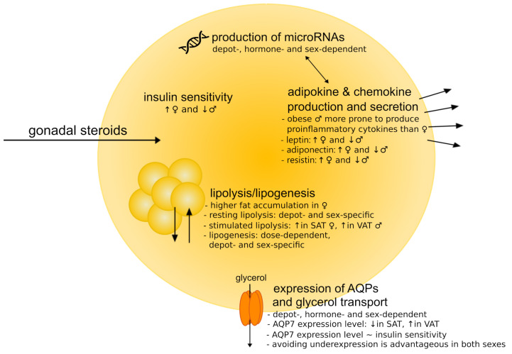
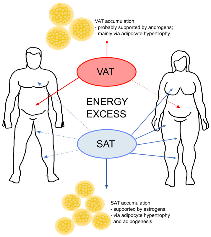
The gonadal steroids, including androgens, estrogens and progestogens, are involved in the control of body fat distribution in humans. Nevertheless, not only the size and localization of the fat depots depend on the sex steroids levels, but they can also highly affect the functioning of adipose tissue. Namely, the gonadocorticoids can directly influence insulin signaling, lipid metabolism, fatty acid uptake and adipokine production. They may also alter energy balance and glucose homeostasis in adipocytes in an indirect way, e.g., by changing the expression level of aquaglyceroporins. This work presents the recent advances in understanding the molecular mechanism of how the gonadal steroids influence the functioning of adipose tissue leading to a set of detrimental metabolic consequences. Special attention is given here to highlighting the sexual dimorphism of adipocyte functioning in terms of health and disease. Particularly, we discuss the molecular background of metabolic disturbances occurring in consequence of hormonal imbalance which is characteristic of some common endocrinopathies such as the polycystic ovary syndrome. From this perspective, we highlight the potential drug targets and the active substances which can be used in personalized sex-specific management of metabolic diseases, in accord with the patient's hormonal status.

Keywords: adipokines; adipose tissue; aquaporins; insulin sensitivity; lipid metabolism; metabolic disorders; microRNA; microRNA-oriented therapy; polycystic ovary syndrome; sex hormones.


Conflict of interest statement

The authors declare no conflict of interest.

Figures

Figure 1
Figure 1
Schematic representation of the most important processes of adipocyte functioning affected by gonadal steroids and the main sex-differences. Sex hormones influence the expression levels of microRNAs, lipolysis and lipogenesis, insulin sensitivity and endocrine function of adipocytes (e.g., adipokine production). One of the most prone connecting links between sex-hormonal imbalance and the disruption of adipose tissue functions is the expression level of aquaporins (AQPs) (in particular, aquaglyceroporins) and, consequently, the effectiveness of the glycerol efflux from adipocyte.
Figure 2
Figure 2
Adipose tissue is an important side for sex hormone interconversions. This figure summarizes the most prominent reactions of this kind. All sex hormones can be synthesized de novo from cholesterol in presence of active steroid hormones. However, more frequently in adipose tissue, the hormones are taken up from plasma and further transformed into other ones. The direction and efficiency within the steroid biosynthetic pathways in adipose tissue depends on the relative expression and activity of steroidogenic enzymes, which are gender-, age- and depot-specific. Abbreviations: CYP11A1, cholesterol side-chain cleavage enzyme; CYP17A1, steroid 17α-monooxygenase; 3β-HSD, 3β-hydroxysteroid dehydrogenase; 17β-HSD, 17β-hydroxysteroid dehydrogenase; SULT2A1, dehydroepiandrosterone sulfotransferase; DHT, dihydrotestosterone.
Figure 3
Figure 3
Schematic representation of sex-dependent differences in the fat deposition patterns. The visceral adipose tissue (VAT) is located around the abdominal organs and is the main type of fat depots in men. On the contrary, women distribute fat mostly in the form of subcutaneous fat. In general, increased visceral mass predominantly results from adipocyte hypertrophy, whereas subcutaneous fat (SAT) grows through both hypertrophy and hyperplasia (with prevailing hyperplastic mechanism) [77,78]. Considering sex-dependent differences in growth of gonadal fat depots, it is observed that, in response to energy excess, male gonadal depot grows mainly through hypertrophy, while female gonadal depots expand both hypertrophy and hyperplasia [79].

Similar articles

Cited by

References

    1. Klaus S. Adipose Tissue. CRC Press; Boca Raton, FL, USA: 2001.
    1. Choe S.S., Huh J.Y., Hwang I.J., Kim J.I., Kim J.B. Adipose tissue remodeling: Its role in energy metabolism and metabolic disorders. Front. Endocrinol. 2016;7:30. doi: 10.3389/fendo.2016.00030. - DOI - PMC - PubMed
    1. Siiteri P.K. Adipose tissue as a source of hormones. Am. J. Clin. Nutr. 1987;45:277–282. doi: 10.1093/ajcn/45.1.277. - DOI - PubMed
    1. Kershaw E.E., Flier J.S. Adipose tissue as an endocrine organ. J. Clin. Endocrinol. Metab. 2004;89:2548–2556. doi: 10.1210/jc.2004-0395. - DOI - PubMed
    1. Gastaldelli A., Gaggini M., DeFronzo R.A. Role of adipose tissue insulin resistance in the natural history of type 2 diabetes: Results from the San Antonio Metabolism Study. Diabetes. 2017;66:815–822. doi: 10.2337/db16-1167. - DOI - PubMed