

Distinct Concentration-Dependent Molecular Pathways Regulate Bone Cell Responses to Cobalt and Chromium Exposure from Joint Replacement Prostheses
- PMID: 34069294
- PMCID: PMC8156984
- DOI: 10.3390/ijms22105225
Distinct Concentration-Dependent Molecular Pathways Regulate Bone Cell Responses to Cobalt and Chromium Exposure from Joint Replacement Prostheses
Abstract
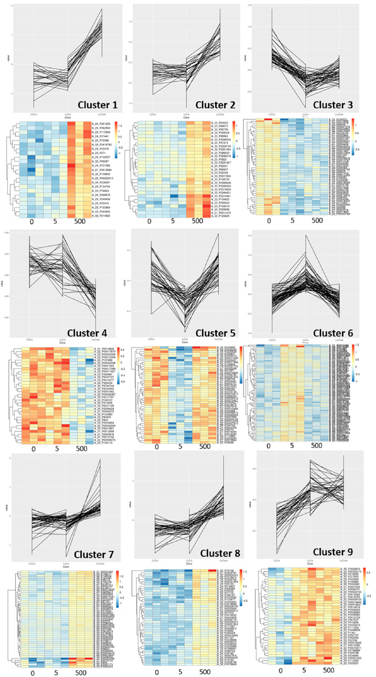
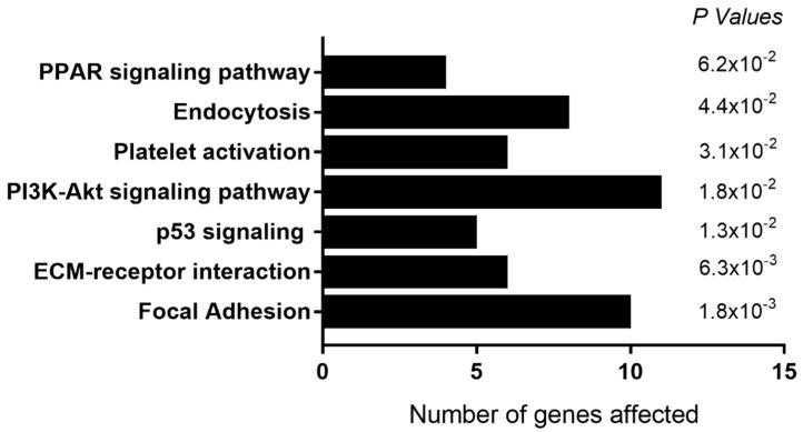
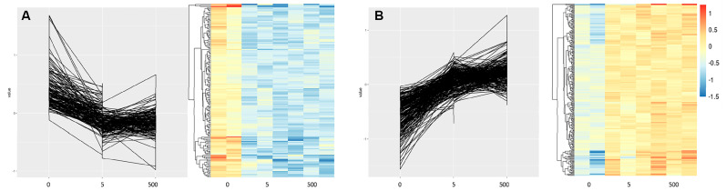
Systemic cobalt (Co) and chromium (Cr) concentrations may be elevated in patients with metal joint replacement prostheses. Several studies have highlighted the detrimental effects of this exposure on bone cells in vitro, but the underlying mechanisms remain unclear. In this study, we use whole-genome microarrays to comprehensively assess gene expression in primary human osteoblasts, osteoclast precursors and mature resorbing osteoclasts following exposure to clinically relevant circulating versus local periprosthetic tissue concentrations of Co2+ and Cr3+ ions and CoCr nanoparticles. We also describe the gene expression response in osteoblasts on routinely used prosthesis surfaces in the presence of metal exposure. Our results suggest that systemic levels of metal exposure have no effect on osteoblasts, and primarily inhibit osteoclast differentiation and function via altering the focal adhesion and extracellular matrix interaction pathways. In contrast, periprosthetic levels of metal exposure inhibit both osteoblast and osteoclast activity by altering HIF-1α signaling and endocytic/cytoskeletal genes respectively, as well as increasing inflammatory signaling with mechanistic implications for adverse reactions to metal debris. Furthermore, we identify gene clusters and KEGG pathways for which the expression correlates with increasing Co2+:Cr3+ concentrations, and has the potential to serve as early markers of metal toxicity. Finally, our study provides a molecular basis for the improved clinical outcomes for hydroxyapatite-coated prostheses that elicit a pro-survival osteogenic gene signature compared to grit-blasted and plasma-sprayed titanium-coated surfaces in the presence of metal exposure.
Keywords: chromium; cobalt; gene expression; hip replacement; microarray; osteoblasts; osteoclasts; prosthesis surface.
Conflict of interest statement
The authors declare no conflict of interest.
Figures
References
-
- Prentice J.R., Clark M.J., Hoggard N., Morton A.C., Tooth C., Paley M.N., Stockley I., Hadjivassiliou M., Wilkinson J.M. Metal-on-Metal Hip Prostheses and Systemic Health: A Cross-Sectional Association Study 8 Years after Implantation. PLoS ONE. 2013;8:e66186. doi: 10.1371/journal.pone.0066186. - DOI - PMC - PubMed
-
- Andrews R.E., Shah K.M., Wilkinson J.M., Gartland A. Effects of cobalt and chromium ions at clinically equivalent concentrations after metal-on-metal hip replacement on human osteoblasts and osteoclasts: Implications for skeletal health. Bone. 2011;49:717–723. doi: 10.1016/j.bone.2011.06.007. - DOI - PubMed
MeSH terms
Substances
Grants and funding
LinkOut - more resources
Full Text Sources
Miscellaneous
