

Machine Learning and Radiomics Applications in Esophageal Cancers Using Non-Invasive Imaging Methods-A Critical Review of Literature
- PMID: 34069367
- PMCID: PMC8158761
- DOI: 10.3390/cancers13102469
Machine Learning and Radiomics Applications in Esophageal Cancers Using Non-Invasive Imaging Methods-A Critical Review of Literature
Abstract
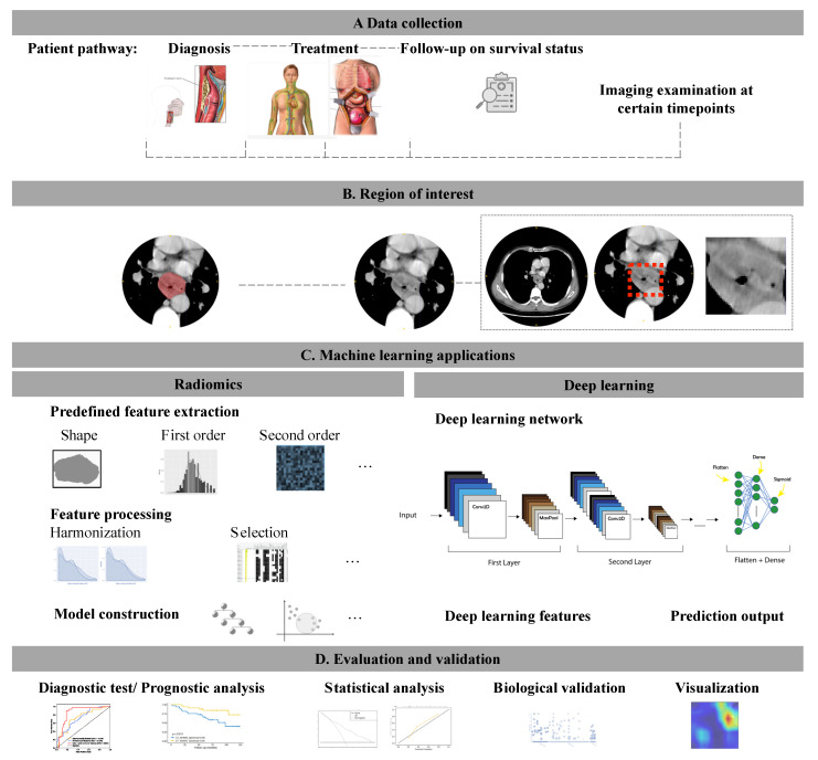
Esophageal cancer (EC) is of public health significance as one of the leading causes of cancer death worldwide. Accurate staging, treatment planning and prognostication in EC patients are of vital importance. Recent advances in machine learning (ML) techniques demonstrate their potential to provide novel quantitative imaging markers in medical imaging. Radiomics approaches that could quantify medical images into high-dimensional data have been shown to improve the imaging-based classification system in characterizing the heterogeneity of primary tumors and lymph nodes in EC patients. In this review, we aim to provide a comprehensive summary of the evidence of the most recent developments in ML application in imaging pertinent to EC patient care. According to the published results, ML models evaluating treatment response and lymph node metastasis achieve reliable predictions, ranging from acceptable to outstanding in their validation groups. Patients stratified by ML models in different risk groups have a significant or borderline significant difference in survival outcomes. Prospective large multi-center studies are suggested to improve the generalizability of ML techniques with standardized imaging protocols and harmonization between different centers.
Keywords: esophageal neoplasms; machine learning; radiology.
Conflict of interest statement
The authors declare no conflict of interest.
Figures
References
-
- Van Hagen P., Hulshof M., Van Lanschot J., Steyerberg E., Henegouwen M.V.B., Wijnhoven B., Richel D., Nieuwenhuijzen G.A., Hospers G.A.P., Bonenkamp J., et al. Preoperative Chemoradiotherapy for Esophageal or Junctional Cancer. N. Engl. J. Med. 2012;366:2074–2084. doi: 10.1056/NEJMoa1112088. - DOI - PubMed
-
- Yang H., Liu H., Chen Y., Zhu C., Fang W., Yu Z., Mao W., Xiang J., Han Y., Chen Z., et al. Neoadjuvant Chemoradiotherapy Followed by Surgery Versus Surgery Alone for Locally Advanced Squamous Cell Carcinoma of the Esophagus (NEOCRTEC5010): A Phase III Multicenter, Randomized, Open-Label Clinical Trial. J. Clin. Oncol. 2018;36:2796–2803. doi: 10.1200/JCO.2018.79.1483. - DOI - PMC - PubMed
-
- Barbetta A., Sihag S., Nobel T., Hsu M., Tan K.S., Bains M., Jones D.R., Molena D. Patterns and risk of recurrence in patients with esophageal cancer with a pathologic complete response after chemoradiotherapy followed by surgery. J. Thorac. Cardiovasc. Surg. 2019;157:1249–1259.e5. doi: 10.1016/j.jtcvs.2018.09.136. - DOI - PMC - PubMed
Publication types
LinkOut - more resources
Full Text Sources
