. 2021 May 19;9(5):1089.
doi: 10.3390/microorganisms9051089.

17-AAG-Induced Activation of the Autophagic Pathway in Leishmania Is Associated with Parasite Death

Affiliations

17-AAG-Induced Activation of the Autophagic Pathway in Leishmania Is Associated with Parasite Death

Antonio Luis de O A Petersen et al. Microorganisms. .

Abstract

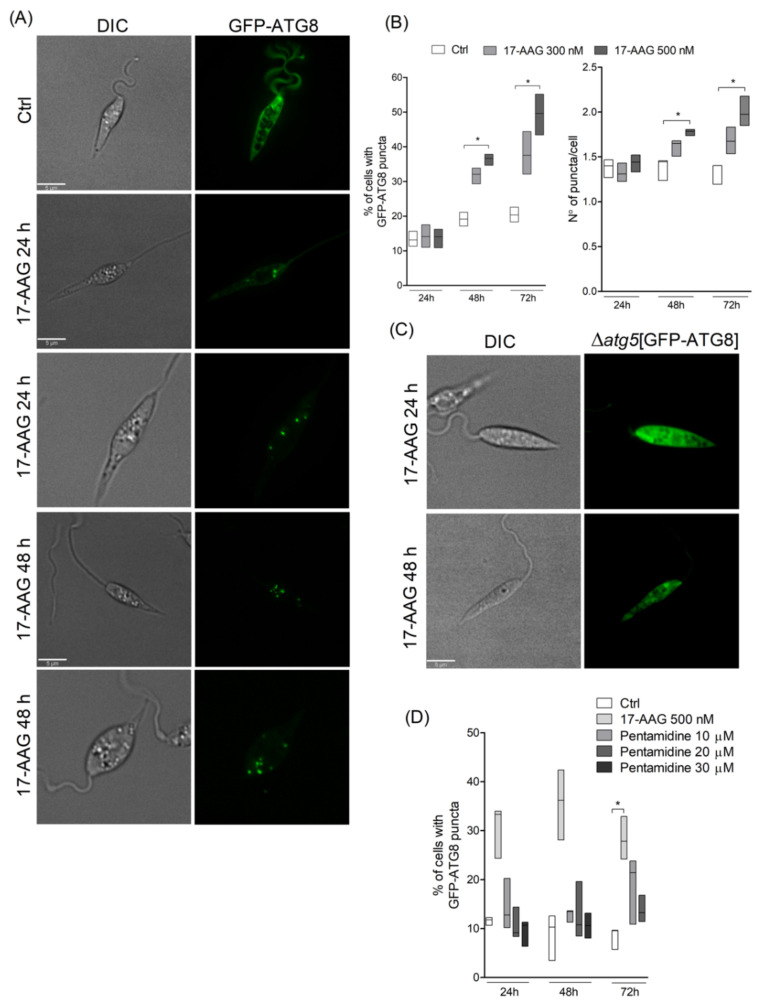
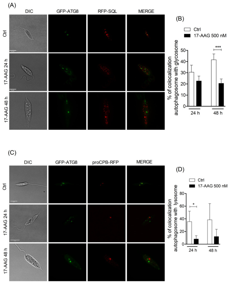
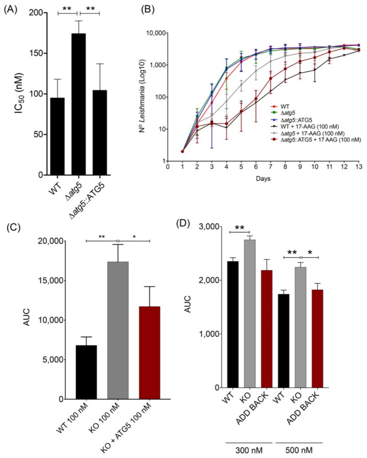
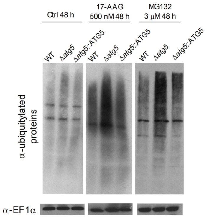
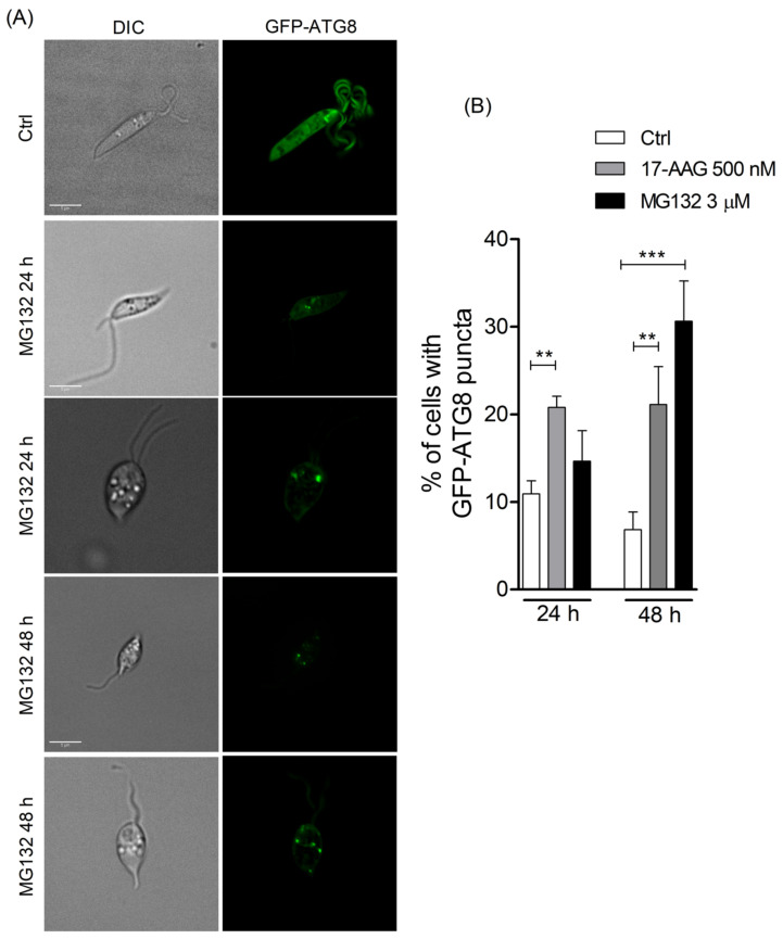
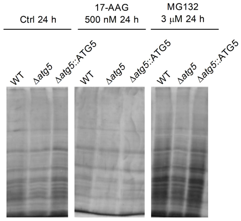
The heat shock protein 90 (Hsp90) is thought to be an excellent drug target against parasitic diseases. The leishmanicidal effect of an Hsp90 inhibitor, 17-N-allylamino-17-demethoxygeldanamycin (17-AAG), was previously demonstrated in both in vitro and in vivo models of cutaneous leishmaniasis. Parasite death was shown to occur in association with severe ultrastructural alterations in Leishmania, suggestive of autophagic activation. We hypothesized that 17-AAG treatment results in the abnormal activation of the autophagic pathway, leading to parasite death. To elucidate this process, experiments were performed using transgenic parasites with GFP-ATG8-labelled autophagosomes. Mutant parasites treated with 17-AAG exhibited autophagosomes that did not entrap cargo, such as glycosomes, or fuse with lysosomes. ATG5-knockout (Δatg5) parasites, which are incapable of forming autophagosomes, demonstrated lower sensitivity to 17-AAG-induced cell death when compared to wild-type (WT) Leishmania, further supporting the role of autophagy in 17-AAG-induced cell death. In addition, Hsp90 inhibition resulted in greater accumulation of ubiquitylated proteins in both WT- and Δatg5-treated parasites compared to controls, in the absence of proteasome overload. In conjunction with previously described ultrastructural alterations, herein we present evidence that treatment with 17-AAG causes abnormal activation of the autophagic pathway, resulting in the formation of immature autophagosomes and, consequently, incidental parasite death.

Keywords: Hsp90; Hsp90 inhibitors; autophagy; chemotherapy; leishmaniasis; ubiquitin.


Conflict of interest statement

The authors declare no conflict of interest. The funders had no role in the design of the study; in the collection, analyses, or interpretation of data; in the writing of the manuscript, or in the decision to publish the results.

Figures

Figure 1
Figure 1
Evaluation of autophagosome formation in Leishmania promastigotes following treatment with 17-AAG. (A) Axenic promastigotes of Leishmania expressing GFP-ATG8 were treated or not with 17-AAG (500 nM) for 24 or 48 h and imaged by fluorescence microscopy. (B) The percentage of cells bearing autophagosomes and the number of autophagosomes per cell were calculated at 24, 48 and 72 h after treatment with 17-AAG (300 or 500 nM). (C) Δatg5[GFP-ATG8] parasites were treated with 17-AAG and imaged by fluorescence microscopy. (D) Comparison of the percentage of cells bearing autophagosomes after treatment with pentamidine (10, 20 or 30 μM) or 17-AAG (500 nM) for 24 and 48 h. Lines within the floating bars represent medians and floating bar quartiles (Q: 25% and 75%) from one out of three independent experiments (Kruskal-Wallis test, Dunn’s multiple comparison test, * p < 0.05).
Figure 2
Figure 2
Analysis of fusion between autophagosomes and glycosomes or lysosomes. (A) Axenic promastigotes of Leishmania expressing GFP-ATG8 and RFP-SQL were treated or not with 17-AAG (500 nM) and imaged by fluorescence microscopy. (B) Quantification of autophagosome-glycosome colocalization after treatment of Leishmania with 17-AAG. (C) Axenic promastigotes of Leishmania expressing ATG8-GFP and proCPB-RFP were treated or not with 17-AAG (500 nM) and imaged by fluorescence microscopy. (D) Quantification of Leishmania autophagosome-lysosome colocalization after treatment with 17-AAG. Bars represent medians ± SD from one out of three independent experiments (Unpaired t test, *** p = 0.0006, * p = 0.0197).
Figure 3
Figure 3
Effect of 17-AAG on survival and replication of WT, Δatg5 and Δatg5::ATG5 Leishmania. (A) IC50 values for 17-AAG in different Leishmania lineages. Bars represent mean ± SD from four independent experiments (One-way ANOVA, Tukey’s multiple comparisons test, ** p < 0.01). (B) Growth curve reflecting 13 day-counts of WT, Δatg5 and Δatg5::ATG5 parasites, treated or not with 17-AAG at 100 nM. Symbols are representative of means ± SD from three independent experiments. (C) Area under the curve (AUC) analysis of WT, Δatg5 and Δatg5::ATG5 growth depicted in panel (B), following treatment with 17-AAG. Bars represent mean ± SD from three independent experiments (one-way ANOVA test, Tukey’s multiple comparison test * p = 0.0321 0.05, ** p = 0.0016). (D) AUC analysis of WT, Δatg5 and Δatg5::ATG5 viability following treatment with 17-AAG at 300 nM and 500 nM for 24 h, 48 h and 72 h. Bars represent mean ± SD of a single experiment performed in quadruplicate (Welch’s ANOVA test, Dunnett’s T3 multiple comparison test * p < 0.05, ** p < 0.01).
Figure 4
Figure 4
Ubiquitylated protein profiles of WT, Δatg5 and Δatg5::ATG5 after 17-AAG or MG132 treatment. WT, Δatg5 and Δatg5::ATG5 parasites were treated with 17-AAG (500 nM) or MG132 (3 µM) for 48 h. Protein extracts were electrophoresed on a 12% gel, blotted and probed with an FK2 anti-ubiquitin antibody. EF1α was used as loading control.
Figure 5
Figure 5
Evaluation of autophagosome formation in promastigotes of Leishmania following treatment with MG132. (A) Promastigotes of Leishmania expressing GFP-ATG8 were treated or not with MG132 (3 μM) and imaged by fluorescence microscopy. (B) The percentage of cells bearing autophagosomes was calculated after treatment with 17-AAG (500 nM) or MG132 (3 μM) for 24 and 48 h. Bars represent mean ± SD of three independent experiments (One-way ANOVA, Tukey’s multiple comparisons test, ** p < 0.01, *** p < 0.001).
Figure 6
Figure 6
Effect of 17-AAG treatment on WT, Δatg5 and Δatg5::ATG5 protein aggregate formation. Culture aliquots from WT, Δatg5 and Δatg5::ATG5 parasites treated with 17-AAG (500 nM) or MG132 (3 µM) for 24 h were withdrawn and analyzed to determine quantities of insoluble protein aggregates by cell lysis and centrifugation. Protein aggregates were subjected to SDS-PAGE followed by silver staining. One experiment is representative of two independent experiments.

References

    1. Herwaldt B.L. Leishmaniasis. Lancet. 1999;354:1191–1199. doi: 10.1016/S0140-6736(98)10178-2. - DOI - PubMed
    1. Banuls A.L., Hide M., Prugnolle F. Leishmania and the leishmaniases: A parasite genetic update and advances in taxonomy, epidemiology and pathogenicity in humans. Adv. Parasitol. 2007;64:1–109. doi: 10.1016/S0065-308X(06)64001-3. - DOI - PubMed
    1. World Health Organization Control of the Leishmaniases. World Health Organ. Tech. Rep. Ser. 2010;Xii–Xiii:1–186. - PubMed
    1. Oliveira L.F., Schubach A.O., Martins M.M., Passos S.L., Oliveira R.V., Marzochi M.C., Andrade C.A. Systematic review of the adverse effects of cutaneous leishmaniasis treatment in the new world. Acta Trop. 2011;118:87–96. doi: 10.1016/j.actatropica.2011.02.007. - DOI - PubMed
    1. Croft S.L., Seifert K., Yardley V. Current scenario of drug development for leishmaniasis. Indian J. Med. Res. 2006;123:399–410. - PubMed

LinkOut - more resources