Case Reports
. 2021 May 19;22(10):5338.
doi: 10.3390/ijms22105338.

A New Mechanism in THRA Resistance: The First Disease-Associated Variant Leading to an Increased Inhibitory Function of THRA2

Affiliations
Case Reports

A New Mechanism in THRA Resistance: The First Disease-Associated Variant Leading to an Increased Inhibitory Function of THRA2

Sarah Paisdzior et al. Int J Mol Sci. .

Abstract

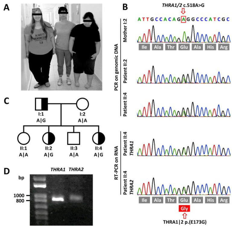
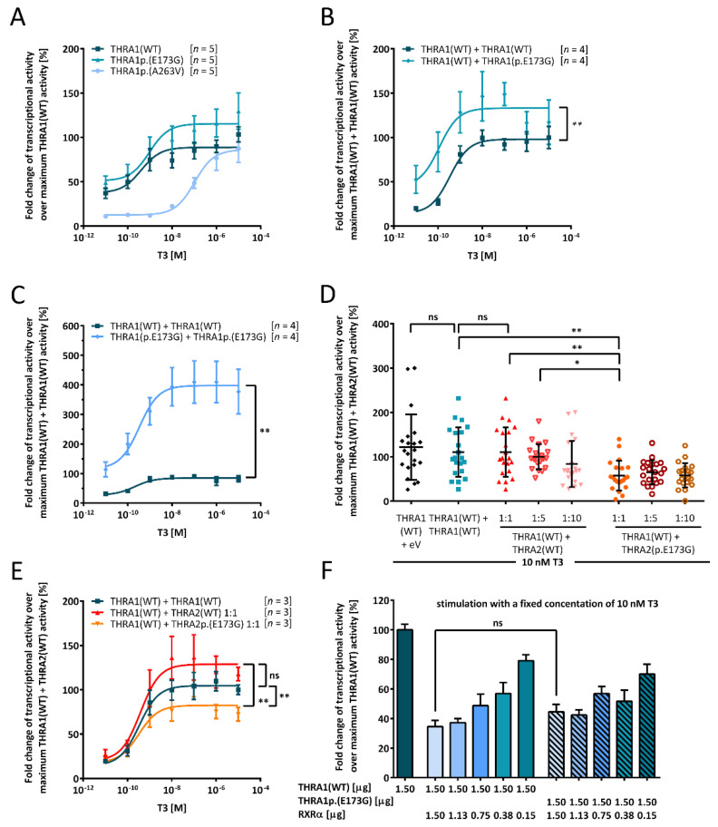
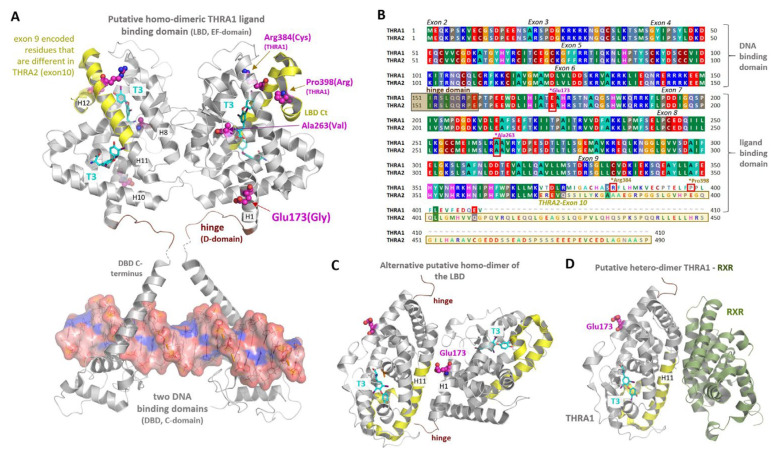
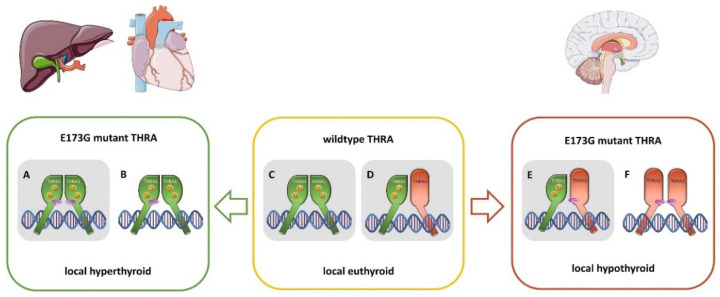
The nuclear thyroid hormone receptors (THRs) are key mediators of thyroid hormone function on the cellular level via modulation of gene expression. Two different genes encode THRs (THRA and THRB), and are pleiotropically involved in development, metabolism, and growth. The THRA1 and THRA2 isoforms, which result from alternative splicing of THRA, differ in their C-terminal ligand-binding domain (LBD). Most published disease-associated THRA variants are located in the LBD of THRA1 and impede triiodothyronine (T3) binding. This keeps the nuclear receptor in an inactive state and inhibits target gene expression. Here, we investigated a new dominant THRA variant (chr17:g.38,241,010A > G, GRCh37.13 | c.518A > G, NM_199334 | p.(E173G), NP_955366), which is located between the DNA- and ligand-binding domains and affects both splicing isoforms. Patients presented partially with hypothyroid (intellectual disability, motor developmental delay, brain atrophy, and constipation) and partially with hyperthyroid symptoms (tachycardia and behavioral abnormalities) to varying degrees. Functional characterization of THRA1p.(E173G) by reporter gene assays revealed increased transcriptional activity in contrast to THRA1(WT), unexpectedly revealing the first gain-of-function mutation found in THRA1. The THRA2 isoform does not bind T3 and antagonizes THRA1 action. Introduction of p.(E173G) into THRA2 increased its inhibitory effect on THRA1, which helps to explain the hypothyroid symptoms seen in our patients. We used protein structure models to investigate possible underlying pathomechanisms of this variant with a gain-of-antagonistic function and suggest that the p.(E173G) variant may have an influence on the dimerization domain of the nuclear receptor.

Keywords: gain-of-antagonistic function; gain-of-function; resistance to thyroid hormones; thyroid hormone receptor alpha.


Conflict of interest statement

The authors declare no conflict of interest.

Figures

Figure 1
Figure 1
Clinical and molecular genetic findings. (A) Photographic image of the mother (I:2) with her affected elder (II:2, left side) and younger (II:4, right side) daughters. (B) Sanger sequencing traces of genomic DNA of both affected sisters and their mother depicting the heterozygous variant in THRA. cDNA sequencing of mRNA extracted from cultured skin fibroblasts of patient II:4 shows that both WT and variant alleles are both expressed albeit at different levels. (C) Pedigree of the family depicting the genotypes at cDNA position c.518. (D) RT-PCR of THRA1 and THRA2 splicing isoforms of patient II:4 shows both isoforms to be expressed. However, as expected [9], in fibroblasts, THRA2 is expressed at lower levels as THRA1.
Figure 2
Figure 2
Functional in vitro characterization of the THRA1p.(E173G) variant. JEG3 cells were co-transfected with T3-responsive luciferase reporter together with the indicated THRA variants: (A) Comparison of THRA1(WT) (dark blue squares), THRA1p.(E173G) (blue triangle), and THRA1p.(A263V) (light blue circles) to investigate the transcriptional effect of the variant. (B) Transfection with either THRA1(WT) (dark blue squares) alone or with the same amount of THRA1(WT) and THRA1p.(E173G) (blue diamonds) to model the patients’ heterozygous state shows a gain-of-function for the variant. Statistical analysis was performed by a Wilcoxon matched-pairs signed rank test. (C) In addition to mimicking the heterozygous state in (B), we also investigated the homozygous state. Here, the amount of DNA encoding for either THRA1(WT) (dark blue squares) or THRA1p.(E173G) (light blue diamonds) was double the amount as in (A). The gain-of-function effect of THRA1p.(E173G) was over-proportionally enhanced, suggesting a gene-dosage effect. For statistical analysis, a Wilcoxon matched-pairs signed-rank test was performed comparing the two datasets. (D) Investigation of the impact of THRA2p.(E173G) on THRA1(WT) revealed a strong antagonistic effect as compared to THRA2(WT) when transfected in a ratio of 1:1. This significant inhibitory effect was also present in comparison to our internal controls of THRA1(WT) + empty vector and of THRA1(WT) + THRA1(WT). Statistical analysis was performed by a non-paired one-way ANOVA comparing individual pairs. (E) Co-expression of THRA1(WT) and either THRA2(WT) (red diamonds) or THRA2p.(E173G) (orange triangles) was performed in a concentration-dependent manner, revealing the enhanced antagonism of THRA2p.(E173G), even with an equal amount of THRA1 and THRA2, which was statistically significant according to a two-way ANOVA followed by Dunnett’s post hoc test. THRA2(WT) did not have a strong effect on THRA1(WT) when expressed in equimolar amounts, since it did not differ from THRA1(WT) (dark blue squares). (F) THRA1p.(E173G) did not have an effect on the formation of heterodimers with RXRα as titration with different amounts of RXRα did not result in a statistical difference to the WT, which was tested by one-way ANOVA with Kruskal–Wallis test performing individual comparison for each expression ratio. For all panels, cells were lysed after stimulation with ascending concentrations of T3 in (A–C,E) or with a fixed concentration of 10 nM T3 in (D,F) for 24 h, and luciferase activity was determined with a luminometer. Values were normalized to the homozygous THRA1/2(WT) state ± SEM for (A–C,E,F), ± SD for (D). (A–C,E) Data resulted from the number of independent experiments provided in the square brackets, each experiment was performed in technical triplicates. (D) Data resulted from seven different independent experiments performed in triplicates. ns, not significant; *, p ≤ 0.05; **, p ≤ 0.01. (F) Data resulted from three different independent experiments.
Figure 3
Figure 3
Structural implications for THRA position Glu173. (A) Two crystal structures (PDB ID: 4lnw) of a monomeric THRA LBD domain (AA 148–406) including the hinge region (brown) with two TH (magenta sticks) binding pockets were superimposed with the dimeric THRB crystal structure (PDB ID: 3d57) to simulate a putative THRA1 LBD dimer constellation. This arrangement in combination with a crystal structure of a dimeric THR:DNA-binding domain (PDB ID: 3m9e) enables the visualization of structural THR features and the potential impact of the p.(E173G) variant. Glu173 is not involved directly in ligand- nor in DNA-binding. In contrast, known pathogenic THRA variants, such as p.(R384C) (THRA1-specific) or p.(A263V) (THRA1- and THRA2-specific) have an impact on the TH-binding process, either by being located inside the TH-binding pocket, or by being linked to protein parts participating in the TH-binding process (e.g., p.(P398R) in the LBD C-terminus, ligand-pocket entrance). In this putative LBD dimer constellation, protomer contacts are mediated by helix 8 (H8) and the transition between H10 and H11. The structural part that would be encoded partially by exon 10 of THRA2 is marked in yellow. (B) The sequence similarities and differences between THRA splicing variants are visualized by the protein sequence alignment. The color-coding distinguishes diverse AA side chain properties, e.g., green for hydrophobic, red for negatively charged, or blue for positively charged. AAs that are encoded by exon 10 (THRA2) are highlighted by a yellow box, which corresponds partially to the yellow backbone-cartoon part in (A). Moreover, exon-encoded sequence dimensions are annotated as well as positions of disease-associated variants shown in (A). (C) An alternative—as compared to (A)—homodimeric THRB constellation (PDB ID: 1n46) was used to simulate a second putative dimeric constellation for the THRA LBD, whereby the Glu173 position would be involved in the dimer-interface. (D) Suggested by the solved THRB/RXR heterodimer complex (PDB ID: 4zo1) and by assuming similar structural properties for THRA and THRB based on very high sequence identity (~75%), Glu173 is likely not involved in the THR/RXR heterodimer-interface.
Figure 4
Figure 4
Schematic overview of the putative THRA dimer constellations in patients with the THRAp.(E173G) variant. In the patient, four different isoforms of THRA [THRA1(WT), THRA1p.(E173G), THRA2(WT), THRA2p.(E173G)] exist and may homo- or hetero-dimerize with each other, depending on their relative abundance in respective cells and organs. This may help to explain the symptoms seen in our patients. THRA1p.(E173G) homodimers (A) and THRA1(WT):THRA1p.(E173G) heterodimers (B), as they are present in peripheral tissues such as cardiomyocytes and hepatocytes, lead to an increased translational activity in these cells, resulting in a local hyperthyroid state as compared to the normally occurring THRA1(WT) homodimer (C). At equimolar concentrations, THRA2(WT) has no antagonistic effect on THRA1(WT) (D). Tissues that predominantly express THRA2, such as in central neurons, THRA2p.(E173G) has an augmented inhibitory effect on THRA1(WT) (E), resulting in a local hypothyroid state in these cells. Other putative heterodimer pairings still have to be investigated, such as THRA2p.(E173G):THRA2p.(E173G) (F), or THRA1p.(E173G):THRA2(WT), and Table 1. p.(E173G):THRA2p.(E173G). In this study, we examined the constellations (A,C,D), and (E), which are highlighted in grey.

Similar articles

Cited by

References

    1. Weinberger C., Thompson C.C., Ong E.S., Lebo R., Gruol D.J., Evans R.M. The C-Erb-A Gene Encodes a Thyroid Hormone Receptor. Nature. 1986;324:641–646. doi: 10.1038/324641a0. - DOI - PubMed
    1. Lazar M.A., Berrodin T.J., Harding H.P. Differential DNA Binding by Monomeric, Homodimeric, and Potentially Heteromeric Forms of the Thyroid Hormone Receptor. Mol. Cell. Biol. 1991;11:5005–5015. doi: 10.1128/MCB.11.10.5005. - DOI - PMC - PubMed
    1. Harvey C.B., Williams G.R. Mechanism of Thyroid Hormone Action. Thyroid Off. J. Am. Thyroid Assoc. 2002;12:441–446. doi: 10.1089/105072502760143791. - DOI - PubMed
    1. Lazar M.A., Hodin R.A., Darling D.S., Chin W.W. Identification of a Rat C-ErbA Alpha-Related Protein Which Binds Deoxyribonucleic Acid but Does Not Bind Thyroid Hormone. Mol. Endocrinol. Baltim. Md. 1988;2:893–901. doi: 10.1210/mend-2-10-893. - DOI - PubMed
    1. Izumo S., Mahdavi V. Thyroid Hormone Receptor Alpha Isoforms Generated by Alternative Splicing Differentially Activate Myosin HC Gene Transcription. Nature. 1988;334:539–542. doi: 10.1038/334539a0. - DOI - PubMed

Publication types

MeSH terms