. 2021 May 19;22(10):5341.
doi: 10.3390/ijms22105341.

Formulative Study and Intracellular Fate Evaluation of Ethosomes and Transethosomes for Vitamin D3 Delivery

Affiliations

Formulative Study and Intracellular Fate Evaluation of Ethosomes and Transethosomes for Vitamin D3 Delivery

Manuela Costanzo et al. Int J Mol Sci. .

Abstract

In this pilot study, ethosomes and transethosomes were investigated as potential delivery systems for cholecalciferol (vitamin D3), whose deficiency has been correlated to many disorders such as dermatological diseases, systemic infections, cancer and sarcopenia. A formulative study on the influence of pharmaceutically acceptable ionic and non-ionic surfactants allowed the preparation of different transethosomes. In vitro cytotoxicity was evaluated in different cell types representative of epithelial, connective and muscle tissue. Then, the selected nanocarriers were further investigated at light and transmission electron microscopy to evaluate their uptake and intracellular fate. Both ethosomes and transethosomes proven to have physicochemical properties optimal for transdermal penetration and efficient vitamin D3 loading; moreover, nanocarriers were easily internalized by all cell types, although they followed distinct intracellular fates: ethosomes persisted for long times inside the cytoplasm, without inducing subcellular alteration, while transethosomes underwent rapid degradation giving rise to an intracellular accumulation of lipids. These basic results provide a solid scientific background to in vivo investigations aimed at exploring the efficacy of vitamin D3 transdermal administration in different experimental and pathological conditions.

Keywords: cell culture; cholecalciferol; cryogenic transmission electron microscopy; in vitro test; light microscopy; lipid nanocarriers; transmission electron microscopy.


Conflict of interest statement

The authors declare no conflict of interest.

Figures
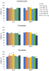
Figure 1
Figure 1
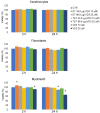
Effect of ET and TET on cell viability of cultured keratinocytes, fibroblasts and myoblasts, as measured by MTT assay. Histograms show the mean percentage value ± s.d. of cell viability after 2 h and 24 h of incubation with ET and TET at different concentrations. CTR: control (untreated) cells. * p < 0.05.
Figure 2
Figure 2
Cryo-TEM images of ET-VD3 (a) and TET-VD3 (b). The bar corresponds to 200 nm in panels (a) and (b), 100 nm in the inset of panel (b).
Figure 3
Figure 3
Effect of ET-VD3, TET-VD3 and VD3 on cell viability of cultured keratinocytes, fibroblasts and myoblasts, as measured by MTT assay. Histograms show the mean percentage value ± s.d. of cell viability after 2 h and 24 h of incubation with nanocarriers or VD3 as in at different concentrations. * p < 0.05.
Figure 4
Figure 4
(a) Mean diameter variation for the indicated ET and TET. Mean diameters were measured by PCS after 0 (yellow) and 3 months (orange) from production and expressed as Z average. Data are the mean of three determinations on different samples; (b) Variation of VD3 content in ET-VD3 (light blue) and TET-VD3 (blue) determined up to 3 months from production. Bars indicate s.d.
Figure 5
Figure 5
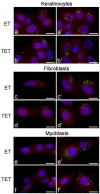
Fluorescence micrographs of keratinocytes, fibroblasts and myoblasts after 2 h (af) and 24 h (a’f’) incubation with ET or TET. Nanocarriers in green (PKH67), cytoplasm in red (trypan blue) and nucleus in blue (Hoechst 33342). Bars 20 μm.
Figure 6
Figure 6
Bright-field micrographs of keratinocytes, fibroblasts and myoblasts after 24 h incubation with ET or TET; CTR are control (untreated) cells. Oil Red O-staining for neutral lipids, hematoxylin counterstaining. Note the marked increase in lipid droplets in TET-treated cells. Bars 20 μm.
Figure 7
Figure 7
Transmission electron micrographs of keratinocytes (a,d,j,k), myoblasts (b,f,g,h) and fibroblasts (c,e,i) after 2 h (ae) and 24 h incubation (fk) with ET. (a) An ET (arrowhead) is entering the cell. (b) A mitochondrion (asterisk) occurs very close to ET distributed in the cytosol. (c) Smooth endoplasmic reticulum into an ET indentation (arrow). (d,e) ET at various degree of degradation: note the smooth vesicles at their periphery (thin arrows) and the decreased electron density (asterisks). (f,g) After 24 h incubation, many ET are distributed in the cytoplasm, sometimes very close to the nucleus (N); note the well-preserved morphology of mitochondria (asterisks), endoplasmic reticulum (arrowheads) and Golgi apparatus (star). (h) Smooth endoplasmic reticulum cisternae surround two ET (arrowheads). (g,h) High magnification details corresponding to the black and white framed areas in (f), respectively. (i,j) Crescent-like ET with smooth endoplasmic reticulum inside their concavity. (k) ET remnants (arrows) surrounded by many smooth vesicles and tubules. Bars 200 nm (ae,gk), 2 μm (f).
Figure 8
Figure 8
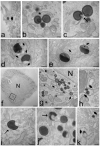
Transmission electron micrographs of keratinocytes (a,f,g,h), fibroblasts (d,e) and myoblasts (b,c,i) and after 2 h (ac) and 24 h incubation (di) with TET. (a) Some TET occur among microvilli on the cell surface. (b) A TET (arrowhead) is entering the cell, while another TET (arrow) occurs free in the cytosol. (c) Some TET are distributed in the cytoplasm, sometimes very close to the nucleus (N); note the crescent-like TET (arrow) and the good morphology of mitochondria (asterisks) and endoplasmic reticulum (arrowheads). (d,e) After 24 h incubation, the cells contain large amounts of lipid droplets (L) of small size; they occur very close each other and are often surrounded by mitochondria (asterisks); (e) high magnification detail corresponding to the framed area in (d). (f) Two coalescing lipid droplets (arrow). (g) After 24 h incubation with TET, some mitochondria swell and lose their cristae (asterisks), and residual bodies (arrows) occur. (h,i) The scarce morphologically recognizable TET show different degradation steps. Bars 200 nm (ac,ei), 1 μm (d).

Similar articles

Cited by

References

    1. Kechichian E., Ezzedine K. Vitamin D and the Skin: An Update for Dermatologists. Am. J. Clin. Dermatol. 2018;19:223–235. doi: 10.1007/s40257-017-0323-8. - DOI - PubMed
    1. Lehmann B., Querings K., Reichrath J. Vitamin D and skin: New aspects for dermatology. Exp. Dermatol. 2004;13:11–55. doi: 10.1111/j.1600-0625.2004.00257.x. - DOI - PubMed
    1. Lehmann B. Role of the vitamin D3 pathway in healthy and diseased skin-facts, contradictions and hypotheses. Exp. Dermatol. 2009;18:97–108. doi: 10.1111/j.1600-0625.2008.00810.x. - DOI - PubMed
    1. Dawson-Hughes B. Vitamin D and muscle function. J. Steroid Biochem. Mol. Biol. 2017;173:313–316. doi: 10.1016/j.jsbmb.2017.03.018. - DOI - PubMed
    1. Abiri B., Vafa M. Vitamin D and Muscle Sarcopenia in Aging. Methods Mol. Biol. 2020;2138:29–47. doi: 10.1007/978-1-0716-0471-7_2. - DOI - PubMed

LinkOut - more resources